Advertisement

Advertisement

Articles

Making Sense of Low Back Pain

Making Sense of Low Back Pain

Teaser: 

Hamilton Hall, MD, FRCSC,1 Julia Alleyne, BHSc(PT), MD, CCFP, Dip. Sport Med MScCH,2 Yoga Raja Rampersaud, MD, FRCSC,3

1Professor, Department of Surgery, University of Toronto; Medical Director, Canadian Back Institute; Executive Director, Canadian Spine Society, Toronto, ON.
2Associate Professor, Department of Family and Community Medicine, University of Toronto, Medical Director, Sport CARE, Women’s College Hospital, Toronto, ON.
3Associate Professor Department of Surgery, University of Toronto, Divisions of Orthopaedic and Neurosurgery, University Health Network Medical Director, Back and Neck Specialty Program, Altum Health, Immediate Past President Canadian Spine Society, Toronto, ON.

CLINICAL TOOLS

Abstract: In 1987, the Quebec Taskforce noted, "Distinct patterns of reliable clinical findings are the only logical basis for back pain categorization and subsequent treatment." Identifying these patterns begins with the patient's history: "Where is your pain the worst?" "Is your pain constant or intermittent?" "Has there been any change in your bowel or bladder function?" This questioning establishes the mechanical nature of the pain, and a physical examination verifies or refutes the pattern established in the history. The examination involves two essential tests to detect upper motor and low sacral root involvement. A failure of the results to fit into one of four syndromes—two back dominant and two leg dominant—suggests a non-mechanical or more complex problem.
Key Words:patterns of back pain, pain location, pain characteristics, history, physical examination.

HealthPlexus is offering an eCME in support of the Back Pain Management Resource

eCME: The Latest in Back Pain Management

This CME activity offers interactive Videos, Animations, Pre- and Post-test Quizzes and you will be able to download a Certificate of Participation upon completion.

90% of Low Back Pain is not related to serious pathology and does not require surgical intervention.
Mechanical Low Back Pain can be categorized to patterns that are identified in history and confirmed in the physical examination.
Findings on radiological imaging including x-ray, CT scan and MRI have not been found to correlate to pain-generating pathology, can increase patient anxiety and detract from successful recovery.
A concise history starts with two questions: "Where is your pain the worst?" and "Is your pain constant or intermittent?"
The goal of physical examination is to verify or refute the diagnostic assumptions made on the basis of the history.
Managing low back pain is not a one-time event. Low back pain is a chronic condition that demands ongoing care and follow-up.
To have access to full article that these tools were developed for, please subscribe. The cost to subscribe is $80 USD per year and you will gain full access to all the premium content on www.healthplexus.net, an educational portal, that hosts 1000s of clinical reviews, case studies, educational visual aids and more as well as within the mobile app.

Managing Back Dominant Pain

Managing Back Dominant Pain

Teaser: 

Hamilton Hall, MD, FRCSC,1 Julia Alleyne, BHSc(PT), MD, CCFP, Dip. Sport Med MScCH,2 Yoga Raja Rampersaud, MD, FRCSC,3

1Professor, Department of Surgery, University of Toronto; Medical Director, Canadian Back Institute; Executive Director, Canadian Spine Society, Toronto, ON.
2Associate Professor, Department of Family and Community Medicine, University of Toronto, Medical Director, Sport CARE, Women’s College Hospital, Toronto, ON.
3Associate Professor Department of Surgery, University of Toronto, Divisions of Orthopaedic and Neurosurgery, University Health Network Medical Director, Back and Neck Specialty Program, Altum Health, Immediate Past President Canadian Spine Society, Toronto, ON.

CLINICAL TOOLS

Abstract: Back dominant pain is either intensified by flexion or is not aggravated by bending forward. The most common pattern, probably discogenic, subdivides into two groups: one with pain on flexion but relief on extension, the other with pain in both directions. The second pattern has symptoms with extension only. Treatment begins with education about the true benign nature of the problem. Mechanical pain responds to posture adjustment and pattern-specific movement. Medication has a secondary role. Imaging is not required for the responding patient. The inability to detect a pattern or a lack of anticipated response combined with non-mechanical findings indicates the need for appropriate referral.
Key Words:back dominant pain, education, medication, imaging, specialist referral.

HealthPlexus is offering an eCME in support of the Back Pain Management Resource

eCME: The Latest in Back Pain Management

This CME activity offers interactive Videos, Animations, Pre- and Post-test Quizzes and you will be able to download a Certificate of Participation upon completion.

Back Dominant pain can be divided into two presentations: pain that is predominantly reproduced with flexion or pain that is reduced or unaffected by flexion.
The recognition of mechanical low back pain is based on a precise history, a validating physical examination and a positive treatment result.
Referred pain to the leg may occur with back dominant pain but, unlike radicular pain, the neurological examination will be normal.
Facilitating the patient to engage in activity that does not aggravate pain is the key to pain management and recovery.
The goal is control, not cure. Anything that relieves the pain and helps to restore mobility is valuable.
Medication has a limited and secondary role. There is no place for the routine use of narcotics or psychotropic drugs.
To have access to full article that these tools were developed for, please subscribe. The cost to subscribe is $80 USD per year and you will gain full access to all the premium content on www.healthplexus.net, an educational portal, that hosts 1000s of clinical reviews, case studies, educational visual aids and more as well as within the mobile app.

Managing Leg Dominant Pain

Managing Leg Dominant Pain

Teaser: 

Yoga Raja Rampersaud, MD, FRCSC,1 Julia Alleyne, BHSc(PT), MD, CCFP, Dip. Sport Med MScCH,2 Hamilton Hall, MD, FRCSC,3

1Associate Professor Department of Surgery, University of Toronto, Divisions of Orthopaedic and Neurosurgery, University Health Network Medical Director, Back and Neck Specialty Program, Altum Health, Immediate Past President Canadian Spine Society, Toronto, ON.
2Associate Professor, Department of Family and Community Medicine, University of Toronto, Medical Director, Sport CARE, Women’s College Hospital, Toronto, ON.
3Professor, Department of Surgery, University of Toronto; Medical Director, Canadian Back Institute; Executive Director, Canadian Spine Society, Toronto, ON.

CLINICAL TOOLS

Abstract: Leg dominant pain suggests direct nerve root involvement: radicular, not referred symptoms. Constant pain associated with positive neurological findings usually results from an acute disc herniation. Symptoms are the result of mechanical compression but principally reflect an inflammatory response, properly designated sciatica. Intermittent leg dominant pain triggered by activity in extension and relieved by rest in flexion probably represents neurogenic claudication: nerve root ischemia secondary to spinal stenosis. Except for acute cauda equina syndrome, acute sciatica is initially managed with scheduled rest, adequate medication, and time. Non-responsive cases may require surgery. Surgery also shows superior outcomes for disabling neurogenic claudication.
Key Words:leg dominant pain, sciatica, neurogenic claudication, cauda equina syndrome, surgery.

HealthPlexus is offering an eCME in support of the Back Pain Management Resource

eCME: The Latest in Back Pain Management

This CME activity offers interactive Videos, Animations, Pre- and Post-test Quizzes and you will be able to download a Certificate of Participation upon completion.

True spine-generated, leg dominant pain is consistently reproduced by particular spinal movements or positions.
No imaging investigation is required for a patient presenting an unequivocal clinical picture and exhibiting steady predictable improvement.
Of the four back pain syndromes, only neurogenic claudication is consistently best treated by surgery.
In contrast to the back dominant cases, in sciatica there is a definite role for short-acting narcotics or psychotropic drugs for uncontrolled pain.
Criteria for Surgical Referral
Emergency Referral The symptoms of Cauda Equina Syndrome are: - Urinary retention followed by insensible urinary overflow. - Unrecognized fecal incontinence. - Loss or decrease in saddle/perineal sensation. Acute Cauda Equina Syndrome is a surgical emergency.
Consider Elective Referral Failure to respond to a trial of conservative care: - Unbearable constant leg dominant pain. - Worsening nerve irritation tests (SLR or femoral nerve stretch). - Expanding motor, sensory or reflex deficits. - Recurrent disabling sciatica. - Disabling neurogenic claudication.
To have access to full article that these tools were developed for, please subscribe. The cost to subscribe is $80 USD per year and you will gain full access to all the premium content on www.healthplexus.net, an educational portal, that hosts 1000s of clinical reviews, case studies, educational visual aids and more as well as within the mobile app.

What's New from the Canadian Consensus Conference on the Diagnosis and Treatment of Dementia (CCCDTD4)

What's New from the Canadian Consensus Conference on the Diagnosis and Treatment of Dementia (CCCDTD4)

Teaser: 

Dr.Michael Gordon 
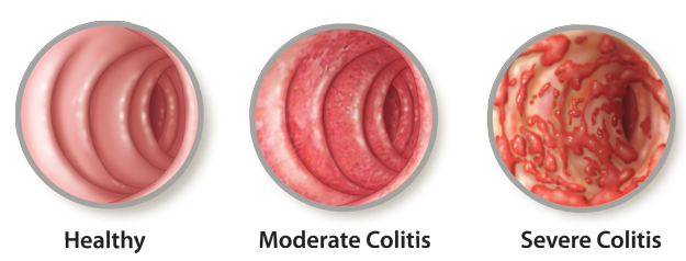
Michael Gordon, MD, MSc, FRCPC, Medical Program Director, Palliative Care, Baycrest Geriatric Health Care System, Professor of Medicine, University of Toronto, Toronto, ON.

In the winter and spring of 2012 I was privileged to be part of the executive working group that organized the fourth Canadian Conference on the Diagnosis and Treatment of Dementia (CCCDTD4) which took place in May 2012. The process as described in both of the initial publications that came out in the fall of 2012 included many participants from all of those physician groups involved in caring for those at risk of dementia, as well as those not-for-profit organizations involved in educating the public.

Comprehensive Patient Care in the Treatment of Ulcerative Colitis

Comprehensive Patient Care in the Treatment of Ulcerative Colitis

Marc Bradette, MD, FRCPC, CSPQ, Clinical Professor, Department of Gastroenterology, Pavillon Hôtel-Dieu de Québec, Québec, QC.


The Patient with Newly Diagnosed Ulcerative Colitis

The Patient with Newly Diagnosed Ulcerative Colitis

Teaser: 

Publication of THE LATEST IN ULCERATIVE COLITIS CARE supplement was made possible by an unrestricted educational grant from Aptalis Pharma

1James Gregor, MD,2Co-authors: John Howard, MD, Nitin Khanna, MD, and Nilesh Chande, MD,

1Division of Gastroenterology, The University of Western Ontario, London, ON.

2are members of the Division of Gastroenterology, London Health Sciences Centre, The University of Western Ontario, London, ON.

CLINICAL TOOLS

Abstract: Informed patients are one of the most important assets available in the management of patients with ulcerative colitis. Clinical experience reinforces that most patients have similar questions upon diagnosis. Anticipating these questions and tailoring them to a particular patient's disease severity and extent should not only streamline follow-up but also mitigate confusion and augment the benefit of the plethora of information available in the 21st century. Using our local experience, we have defined the 10 most common questions asked by patients and modified the answers, where necessary, to improve their specificity to patients with ulcerative proctitis, left-sided ulcerative colitis, and pancolitis.
Key Words: ulcerative colitis, patient, questions, classification, management.

Patients can be relatively ill informed regarding the nature of their UC, its management, and its ultimate prognosis.
Generally, disease extent is divided into three categories: ulcerative proctitis, left-sided disease, and pancolitis.
A simple approach with frequently asked questions (FAQs) is a highly desirable and efficient means of transmitting information.
Clinical experience reinforces that most patients have similar questions upon diagnosis with UC.
Anticipating these questions and tailoring them to a particular patient's disease severity and extent should streamline follow-up and also mitigate confusion.
To have access to full article that these tools were developed for, please subscribe. The cost to subscribe is $80 USD per year and you will gain full access to all the premium content on www.healthplexus.net, an educational portal, that hosts 1000s of clinical reviews, case studies, educational visual aids and more as well as within the mobile app.

Phthalates in 5-Aminosalicylates: Informing Therapeutic Choice and Minimizing Risk

Phthalates in 5-Aminosalicylates: Informing Therapeutic Choice and Minimizing Risk

Teaser: 

Publication of THE LATEST IN ULCERATIVE COLITIS CARE supplement was made possible by an unrestricted educational grant from Aptalis Pharma

Geoffrey C. Nguyen, MD, PhD,

Associate Professor of Medicine, Mount Sinai Hospital Centre for Inflammatory Bowel Disease, University of Toronto, Toronto, ON.

CLINICAL TOOLS

Abstract: 5-Aminosalicylates (5-ASAs) are considered first-line therapy for mild to moderate ulcerative colitis because of their proven effectiveness and safety profile, even in pregnancy. One formulation, however, contains dibutyl phthalate (DBP) in its coating. Though DBP may cause disruptions in utero reproductive development and other congenital abnormalities in rodents, it is unclear whether it leads to physiologically significant fetal abnormalities in humans. The US Food and Drug Administration has changed its classification for DBP-containing 5-ASAs from pregnancy category B to pregnancy category C to reflect a greater degree of uncertainty regarding its effect in humans. For pregnant women with ulcerative colitis, the most important message is to take their inflammatory bowel disease (IBD) medications to prevent disease relapse, which may have the most adverse effects on pregnancy. Physicians should, however, discuss with young women who are taking 5-ASA with DBP the benefits and risks of switching to another formulation of 5-ASA without the DBP compound.
Key Words: phthalates, 5-aminosalicylate, ulcerative colitis, dibutyl phthalate, pregnancy.

5-Aminosalicylates (5-ASAs) are effective for the treatment of mild to moderate ulcerative colitis and are generally regarded as safe to use, even during pregnancy.
Dibutyl phthalate (DBP) is found in the coating of certain formulations of 5-ASA, and in rodents has been shown to be associated with developmental and congenital abnormalities.
Though phthalates have been shown to be associated with some indicators of reduced masculinization among male fetuses, there is insufficient evidence to prove that it leads to significant harmful effects.
There are several formulations of 5-ASA that do not contain DBP.
Asacol, which contains DBP, is categorized as a pregnancy category C drug, while most other 5-ASAs are in pregnancy category B.
It should be emphasized to pregnant women that taking medications for their inflammatory bowel disease is important because the disease has a strong impact on, not just their health, but the health of their fetus too.
Women of child-bearing age who are taking a DBP-containing 5-ASA should have a discussion regarding the benefits and risks of switching to another 5-ASA, preferably before pregnancy.
To have access to full article that these tools were developed for, please subscribe. The cost to subscribe is $80 USD per year and you will gain full access to all the premium content on www.healthplexus.net, an educational portal, that hosts 1000s of clinical reviews, case studies, educational visual aids and more as well as within the mobile app.

Ulcerative Colitis: A Case Study

Ulcerative Colitis: A Case Study

Teaser: 

Publication of THE LATEST IN ULCERATIVE COLITIS CARE supplement was made possible by an unrestricted educational grant from Aptalis Pharma

Brian Bressler, MD, MS, FRCPC,

Clinical Assistant Professor of Medicine, Division of Gastroenterology, St. Paul's Hospital, University of British Columbia, Vancouver, BC.

CLINICAL TOOLS

Abstract: A 28-year-old male presented to our office for a consultation about his bloody bowel movements. Colonoscopy revealed moderately active left-sided ulcerative colitis extending from the anal verge up to the mid-descending colon. He was prescribed both oral and rectal 5-ASAs for induction therapy, and is in remission. Appropriate patient education has helped him realize that the best chance of staying in remission is to continue on his medical therapy.
Key Words: ulcerative colitis, 5-aminosalicylate, medication adherence, dysplasia surveillance, rectal inflammation.

Stool samples should be tested for infectious causes of bloody diarrhea.
Treatment with steroids should be avoided, if possible, as this medication carries the most risk.
In most cases, clinical remission is an acceptable outcome.
In patients newly diagnosed with left-sided ulcerative colitis, if macroscopic evidence of inflammation stops before 35 cm from the anal verge, it is critical to take biopsies in the proximal left colon in normal-appearing mucosa to determine whether a patient with left-sided disease will require dysplasia surveillance.
Patient education at each follow-up visit helps to ensure medication adherence.
We need to help patients understand that UC can be managed with medication, but not cured.
To have access to full article that these tools were developed for, please subscribe. The cost to subscribe is $80 USD per year and you will gain full access to all the premium content on www.healthplexus.net, an educational portal, that hosts 1000s of clinical reviews, case studies, educational visual aids and more as well as within the mobile app.

Optimizing Targets in Patient Management of Ulcerative Colitis: The Role of Fecal Calprotectin in Guiding Maintenance Therapy

Optimizing Targets in Patient Management of Ulcerative Colitis: The Role of Fecal Calprotectin in Guiding Maintenance Therapy

Teaser: 

Publication of THE LATEST IN ULCERATIVE COLITIS CARE supplement was made possible by an unrestricted educational grant from Aptalis Pharma

A. Hillary Steinhart, MD,

Member of the Division of Gastroenterology, Mount Sinai Hospital/University Health Network, Professor of Medicine, University of Toronto, Toronto, ON.

CLINICAL TOOLS

Abstract: Although medical therapy for ulcerative colitis is usually effective at inducing clinical remission, numerous studies have shown that patients in clinical remission may have ongoing and varying degrees of mucosal inflammation. It appears that patients who have greater degrees of active mucosal inflammation, despite the absence of clinical symptoms, are at higher risk of developing a symptomatic flare in the near term. In patients with UC, the level of calprotectin in stool correlates not only with the degree of clinical severity but also with the presence or absence of mucosal inflammation. These findings raise the possibility of using fecal calprotectin as a non-invasive means of monitoring patients in clinical remission and adjusting treatment in those who demonstrate a rise in fecal calprotectin, before symptoms recur.
Key Words: ulcerative colitis, fecal calprotectin, flare prediction, mucosal inflammation, non-invasive monitoring.

Patients who experience a symptomatic flare after having been in clinical remission often have increased mucosal inflammation that predates the flare—sometimes by several months.
With the importance of mucosal healing acknowledged, there has been increasing interest in more frequent assessment of mucosal healing and mucosal inflammation.
This has led to the examination of a number of non-invasive and less expensive means of assessing these parameters.
The presumption is that if such risk factors can be identified, then effective interventions can be applied earlier in the course of disease in order to prevent a clinical flare.
In patients with UC, the level of fecal calprotectin correlates not only with the degree of clinical severity but also with the presence of absence of mucosal inflammation.
These findings raise the possibility of using fecal calprotectin as a non-invasive means of monitoring patients in clinical remission, and adjusting treatment in those who demonstrate a rise in fecal calprotectin, before symptoms recur.
To have access to full article that these tools were developed for, please subscribe. The cost to subscribe is $80 USD per year and you will gain full access to all the premium content on www.healthplexus.net, an educational portal, that hosts 1000s of clinical reviews, case studies, educational visual aids and more as well as within the mobile app.