Advertisement

Advertisement

cauda equina syndrome

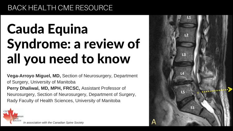
Cauda Equina Syndrome: a review of all you need to know

Teaser: 

Vega-Arroyo Miguel, MD,1 Perry Dhaliwal, MD, MPH, FRCSC,2

1 Section of Neurosurgery, Department of Surgery, University of Manitoba.
2 Assistant Professor of Neurosurgery, Section of Neurosurgery, Department of Surgery, Rady Faculty of Health Sciences, University of Manitoba.

CLINICAL TOOLS

Abstract: Cauda equina syndrome (CES) is the collection of signs and symptoms produced by severe compression of the lumbar spinal nerves that form the cauda equina. The compression can be caused by lumbar degenerative changes, intraspinal tumors, epidural hematoma, and infections. Rapid diagnosis and treatment are paramount as CES requires emergent surgical decompression. With delay, the patient could develop permanent neurological deficits including loss of lower limb sensorimotor function, bladder, bowel, and/or sexual dysfunction. Unfortunately, even with expeditious surgery, neurological improvements remain unpredictable. Failure to fully explain the possible prognoses can involve all the healthcare providers in medicolegal consequences.
Key Words:Cauda Equina Syndrome, Spine Emergency, Urinary retention, MRI scanning, Saddle Anesthesia.

Members of the College of Family Physicians of Canada may claim MAINPRO-M2 Credits for this unaccredited educational program.

www.cfpc.ca/Mainpro_M2

You can take quizzes without subscribing; however, your results will not be stored. Subscribers will have access to their quiz results for future reference.

1. Cauda Equina Syndrome results from pathologies that compress the nerves in the lumbosacral spinal canal, most commonly due to an acute lumbar disc herniation.
2. Early diagnosis is crucial and is made clinically by distinctive symptoms of saddle anesthesia, acute urinary incontinence combined with acute back and leg pain.
3. The most consistent early clinical sign of CES is urinary retention, and the prognosis is worse when present.
4. Urgent MRI is the study of choice and should be performed to confirm or rule out CES.
5. Surgery is highly recommended within 24 hours after CES is identified.
1. Cauda Equina Syndrome is caused by a large space-occupying lesion within the central canal of the lumbosacral spine, most commonly a large disc herniation. However, compression can also be caused by lumbar degenerative changes, intraspinal tumors, epidural hematoma, and infections.
2. Cauda equina syndrome generally presents with varying degrees of sensory loss and motor weakness in the lower extremities, saddle anesthesia, and bowel/bladder dysfunction (these last 2 are required to establish the diagnosis of CES).
3. The main clinical feature between differentiating Cauda Equina Syndrome vs Conus Medullaris Syndrome, is the absence of UPPER MOTOR NEURONS findings in CES).
4. About 70% of patients with cauda equina syndrome have a previous history of lower back pain and/or sciatica.
5. Although the prognosis is largely determined by the preoperative severity of neurological deficits, early surgery improves the chance of significant recovery so patients with CES require urgent surgical intervention.
To have access to full article that these tools were developed for, please subscribe. The cost to subscribe is $80 USD per year and you will gain full access to all the premium content on www.healthplexus.net, an educational portal, that hosts 1000s of clinical reviews, case studies, educational visual aids and more as well as within the mobile app.

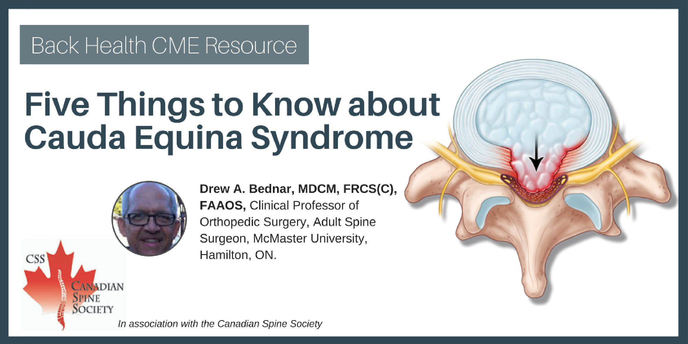
Five Things to Know about Cauda Equina Syndrome

Teaser: 

Drew A. Bednar, MDCM, FRCS(C), FAAOS,

Clinical Professor of Orthopedic Surgery, Adult Spine Surgeon, McMaster University, Hamilton, ON.

CLINICAL TOOLS

Abstract: Cauda Equina Syndrome (CES) is a rare progressive syndrome of pain and neurological deficits below the waist caused by massive central lumbar disc prolapse. The most common clinical presentation is highly variable with multifocal mixed polyradicular deficits. Loss of bladder and/or bowel control can be subtle and is frequently not the patient' chief complaint. These symptoms must be aggressively sought by the assessing physician. While delays of a few hours in the diagnosis and management may not be deleterious, definitive lumbar MRI imaging and (if positive) surgical care referral are emergent.
Key Words: Cauda Equina Syndrome; Presentation; Diagnosis; Outcome.

Members of the College of Family Physicians of Canada may claim MAINPRO-M2 Credits for this unaccredited educational program.

www.cfpc.ca/Mainpro_M2

You can take quizzes without subscribing; however, your results will not be stored. Subscribers will have access to their quiz results for future reference.

The CES patient often presents with rapidly escalating, poorly controlled pain. There may be daily or even more frequent physician visits.
While standing, the CES patient commonly adopts a "sciatic scoliosis": forward bending at the waist and leaning to either side as they attempt to decompress themselves. They may limp or need walking aids. These features help distinguish them from drug-seekers or malingerers.
Since patients are distracted by extreme pain, they may not reliably volunteer a history of bladder/bowel disturbance.
Cauda Equina Syndrome is an acute or subacute pathology caused specifically by massive central prolapse of a lumbar disc. Decompensation lumbar spinal stenosis is not Cauda Equina Syndrome.
Cauda Equina Syndrome most commonly presents with complaints of back or leg pain. These differ from common sciatica in being rapidly progressive, difficult to control with analgesics and often associated with considerable locomotor impairment.
The neurological examination in Cauda Equina Syndrome most commonly finds a mixed pattern of incomplete polyradicular deficits in the distribution of multiple lumbar and sacral nerve roots involving either of the legs and/or the saddle (perineum). The classically described complete flaccidity with loss of all motor control from the waist down is extremely rare.
Patients presenting with CES will not commonly volunteer complaints of incontinence or urinary retention as they are often overwhelmed by the magnitude of their pain. The assessor must specifically ask about bowel/bladder function and when indicated, test these by bladder scanning or catheterizing and a digital rectal examination.
As a rapidly evolving syndrome of neurological deterioration, CES warrants emergent imaging investigation and referral. Although the literature is not precise on the critical time point, it is widely accepted that patients should receive surgical intervention within 24 to 48 hours.
To have access to full article that these tools were developed for, please subscribe. The cost to subscribe is $80 USD per year and you will gain full access to all the premium content on www.healthplexus.net, an educational portal, that hosts 1000s of clinical reviews, case studies, educational visual aids and more as well as within the mobile app.
Disclaimer: 
Disclaimer at the end of each page

Managing Leg Dominant Pain

Managing Leg Dominant Pain

Teaser: 

Yoga Raja Rampersaud, MD, FRCSC,1 Julia Alleyne, BHSc(PT), MD, CCFP, Dip. Sport Med MScCH,2 Hamilton Hall, MD, FRCSC,3

1Associate Professor Department of Surgery, University of Toronto, Divisions of Orthopaedic and Neurosurgery, University Health Network Medical Director, Back and Neck Specialty Program, Altum Health, Immediate Past President Canadian Spine Society, Toronto, ON.
2Associate Professor, Department of Family and Community Medicine, University of Toronto, Medical Director, Sport CARE, Women’s College Hospital, Toronto, ON.
3Professor, Department of Surgery, University of Toronto; Medical Director, Canadian Back Institute; Executive Director, Canadian Spine Society, Toronto, ON.
CLINICAL TOOLS

Abstract: Leg dominant pain suggests direct nerve root involvement: radicular, not referred symptoms. Constant pain associated with positive neurological findings usually results from an acute disc herniation. Symptoms are the result of mechanical compression but principally reflect an inflammatory response, properly designated sciatica. Intermittent leg dominant pain triggered by activity in extension and relieved by rest in flexion probably represents neurogenic claudication: nerve root ischemia secondary to spinal stenosis. Except for acute cauda equina syndrome, acute sciatica is initially managed with scheduled rest, adequate medication, and time. Non-responsive cases may require surgery. Surgery also shows superior outcomes for disabling neurogenic claudication.
Key Words:leg dominant pain, sciatica, neurogenic claudication, cauda equina syndrome, surgery.

HealthPlexus is offering an eCME in support of the Back Pain Management Resource

eCME: The Latest in Back Pain Management

This CME activity offers interactive Videos, Animations, Pre- and Post-test Quizzes and you will be able to download a Certificate of Participation upon completion.

True spine-generated, leg dominant pain is consistently reproduced by particular spinal movements or positions.
No imaging investigation is required for a patient presenting an unequivocal clinical picture and exhibiting steady predictable improvement.
Of the four back pain syndromes, only neurogenic claudication is consistently best treated by surgery.
In contrast to the back dominant cases, in sciatica there is a definite role for short-acting narcotics or psychotropic drugs for uncontrolled pain.
Criteria for Surgical Referral
Emergency Referral The symptoms of Cauda Equina Syndrome are: - Urinary retention followed by insensible urinary overflow. - Unrecognized fecal incontinence. - Loss or decrease in saddle/perineal sensation. Acute Cauda Equina Syndrome is a surgical emergency.
Consider Elective Referral Failure to respond to a trial of conservative care: - Unbearable constant leg dominant pain. - Worsening nerve irritation tests (SLR or femoral nerve stretch). - Expanding motor, sensory or reflex deficits. - Recurrent disabling sciatica. - Disabling neurogenic claudication.
To have access to full article that these tools were developed for, please subscribe. The cost to subscribe is $80 USD per year and you will gain full access to all the premium content on www.healthplexus.net, an educational portal, that hosts 1000s of clinical reviews, case studies, educational visual aids and more as well as within the mobile app.