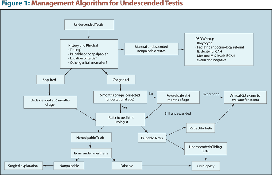
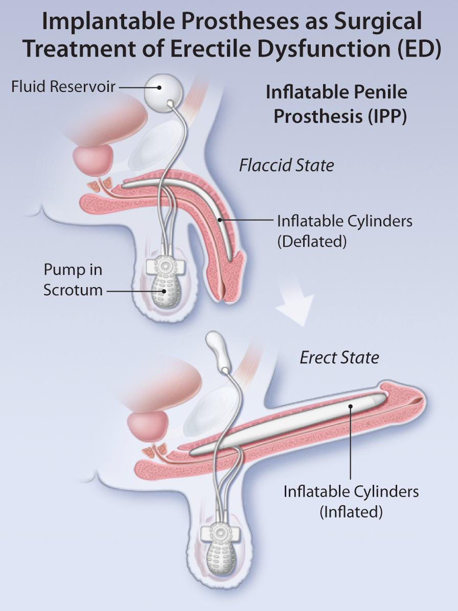
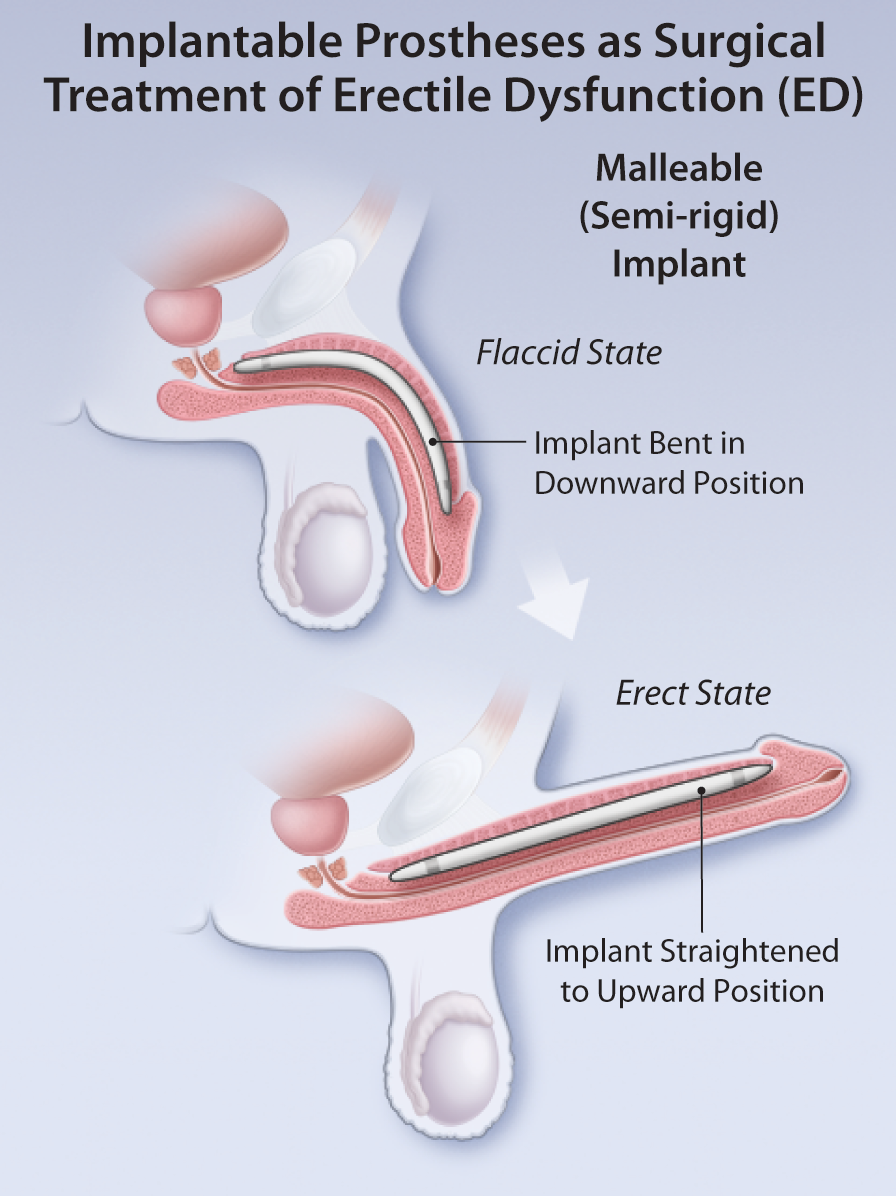
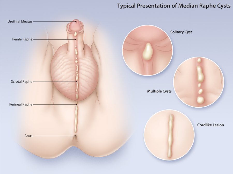
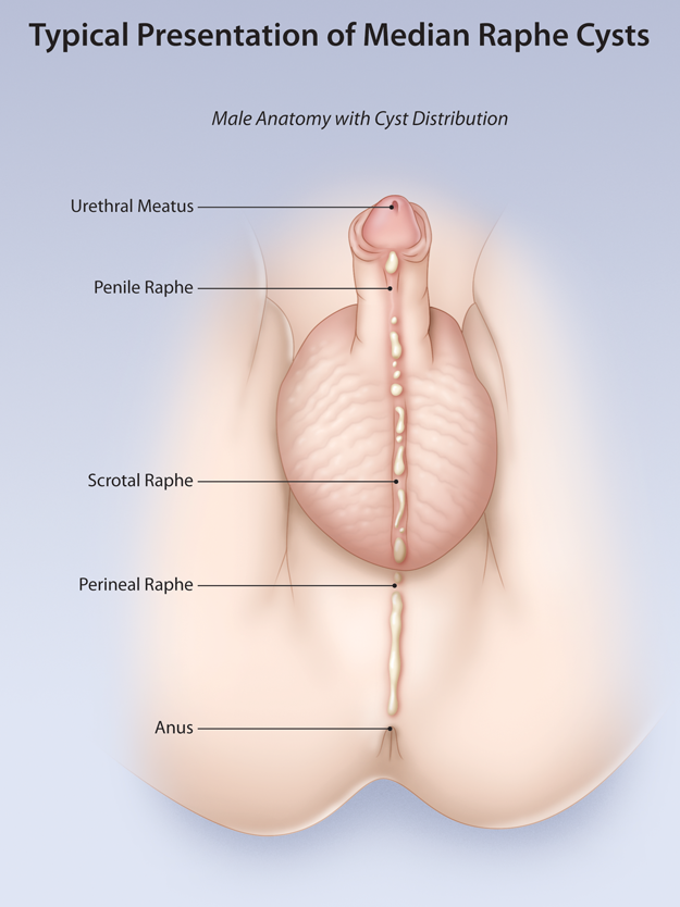
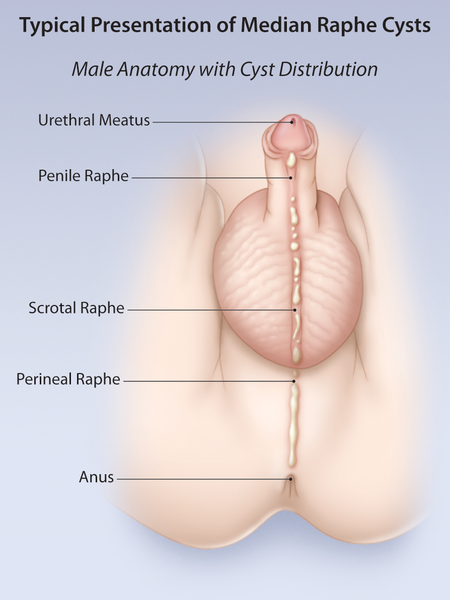
Advertisement

Advertisement

Articles

Connecting the Spots: Hyperpigmented Lesions in Children

Teaser: 

Lisa M. Flegel,1 Joseph M. Lam, MD, FRCSC,2

1Medical Degree Undergraduate Program, Northern Medical Program, University of British Columbia, BC.
2Clinical Assistant Professor, Department of Pediatrics and Dermatology, University of British Columbia, BC.

CLINICAL TOOLS

Abstract: Hyperpigmented lesions are common in the pediatric population and identifying their etiologies can be challenging for physicians. Patients and caregivers may worry that hyperpigmented lesions are dangerous, associated with an internal illness or that they may lead to skin cancers. Having a better understanding of the causes and natural histories of these lesions may help to guide management and alleviate worry. This review article will provide an overview of select common and uncommon causes of hyperpigmented skin lesions in children.
Key Words: hyperpigmentation, pediatric.
1. Most hyperpigmented lesions in children do not require treatment aside from for cosmesis.
2. Features of malignant melanoma in children include: non-pigmented, uniform color, variable diameter, nodular lesions, and occurring de novo.
3. Parents and children should be warned that melanocytic nevi will grow as their child grows, but growth should be proportionate.
4. The risk of melanocytic nevi becoming malignant melanoma in children is very small.
In children with numerous melanocytic nevi, a good rule of thumb is to look for the 'ugly duckling' mole.
To track lesions over time, parents can develop a routine of taking a picture each year on the child's birthday.
To have access to full article that these tools were developed for, please subscribe. The cost to subscribe is $80 USD per year and you will gain full access to all the premium content on www.healthplexus.net, an educational portal, that hosts 1000s of clinical reviews, case studies, educational visual aids and more as well as within the mobile app.

Interventional Radiology Procedures for Chronic Low Back Pain

Teaser: 

Dr. Ted Findlay, D.O., CCFP,1 Amar Suchak, MD, FRCP(C), 2

1is a Clinical Assistant Professor in the Department of Medicine at the University of Calgary. He is also in a Private Family Medicine practice. In addition he is on Medical Staff at Alberta Health Services, Calgary Zone in Calgary, Alberta.
2Clinical Assistant Professor Department of Radiology, Department of Nuclear Medicine, University of Calgary, Calgary, Alberta.

CLINICAL TOOLS

Abstract: There is an increasing availability and clinical use of interventional radiological techniques for patients with low back pain. This can be a valuable additional tool in the management of low back pain that has not responded to conservative treatment. However, the clinical indications and appropriate uses as well as cautions that apply to this treatment modality are in many cases less well understood by the primary care practitioner. The objective of this article is to review clinical scenarios in which these procedures are commonly considered, as well as their limitations. The field of interventional radiology is one that is rapidly evolving and an area of active clinical research. It is important for the primary care practitioner to have a basic understanding of the current state of the art in order to have an informed discussion with their patients who may be seeking advice on this treatment option.
Key Words: Low back pain; treatment; interventional radiology definitions; interventional radiology indications; interventional radiology complications.

Members of the College of Family Physicians of Canada may claim MAINPRO-M2 Credits for this unaccredited educational program.

www.cfpc.ca/Mainpro_M2

You can take quizzes without subscribing; however, your results will not be stored. Subscribers will have access to their quiz results for future reference.

1. In patients carefully selected by clinical and radiological examination, there can be satisfying clinical gains from the use of currently available interventional radiologic procedures.
2. One must not assume that abnormal findings on radiologic imaging immediately explains the anatomical cause of a patient's low back pain; a corresponding accurate history and physical examination is ideal prior to commencing injections.
3. When successful, the gains from radiological interventions should be considered one portion of a broader clinical treatment plan, rather than the entire plan of management.
4. Unsuccessful interventional procedures should not be repeated.
1. Do not apply repeated interventional procedures with an expectation that one of them will find the target source of the patient's low back pain.
2. Although they may be uncommon, interventional radiology risks can occur and the referring physician should be cognizant of these dangers that accumulate with repeated interventions.
To have access to full article that these tools were developed for, please subscribe. The cost to subscribe is $80 USD per year and you will gain full access to all the premium content on www.healthplexus.net, an educational portal, that hosts 1000s of clinical reviews, case studies, educational visual aids and more as well as within the mobile app.

A New Approach for the Excision of a Vallecular Cyst Using a Zero-Degree Nasal Endoscope

Teaser: 

Dr. Pradeep Shenoy, MD, FRCS, FACS, DLO,

ENT & Neck Surgeon, Campbellton Regional Hospital, Campbellton, New Brunswick, Canada.

CLINICAL TOOLS

Abstract: This case study reviews the clinical presentation and treatment of a patient’s vascular cyst. Though it is a rare diagnosis/condition, it could be a medical emergency for an individual of any age. Described here is a new approach for the complete excision of the vallecular cyst.
Key Words: vallecular cyst, excision, zero-degree nasal endoscope
Vallecular cysts are retention cysts in the Vallecular—a space between the base of the tongue, epiglottis and lateral pharyngeal wall.
They are triggered by acid reflux and smoking.
Vallecular cyst can be seen in CT Scan and laryngoscopy examination.
The access is difficult trans-orally. Here we are describing New approach using a zero-degree endoscope.
Vallecular cysts are rare in the paediatric and adult age groups.
Vallecular cysts can present as asymptomatic when small, however, when big they can present as a feeling of some food stuck in the throat or pain.
In emergency situation can block the food and airway passage and require emergency treatment.
To have access to full article that these tools were developed for, please subscribe. The cost to subscribe is $80 USD per year and you will gain full access to all the premium content on www.healthplexus.net, an educational portal, that hosts 1000s of clinical reviews, case studies, educational visual aids and more as well as within the mobile app.

About Psoriasis

About Psoriasis

WHAT IS PSORIASIS?

Psoriasis is a common but chronic skin condition that causes inflammation and scaling (red elevated patches and flaking silvery scales). The patches can be itchy or sore, causing discomfort and pain. Psoriasis causes skin cells to rise to the surface and shed at a very rapid rate. On average, people with psoriasis shed their skin cells every 3 to 4 days, while people without the condition have a turnover rate of about every 30 days.1,2,3,4

Median Raphe Cysts

Teaser: 

Mary Tong, BHSc, MD Candidate,1 Joseph M. Lam, MD, FRCSC,2

1McMaster University, Hamilton, ON.
2Clinical Associate Professor, Department of Pediatrics, Clinical Associate Professor, Department of Dermatology University of British Columbia, BC.

CLINICAL TOOLS

Abstract: Median raphe cysts are rare congenital lesions caused by a defect in embryological development of the male genitalia. They can present as solitary or multiple papules along the median raphe from urethral meatus to the anus. Although they are asymptomatic during childhood, they can cause problems later on as they increase in size. Surgical excision of the lesion is not necessary unless the patient becomes symptomatic.
Key Words: median raphe cysts, congenital lesions, treatment, management.
Median raphe cysts are benign cysts that can be present at birth, or acquired due to trauma or infection in the genitalia area.
Histologically, the cysts can have pseudo stratified columnar, squamous cell, or glandular epithelium, or a mixture of these cells.
Although these cysts are asymptomatic during childhood, they should be monitored overtime because they may cause problems as they increase in size with time.
Because these are benign malformations, median raphe cysts do not require excision unless they cause problems such as pain, problems with urination or sexual activity, or for cosmetic reasons.
Median raphe cysts are benign lesions that may be caused be a defect in the embryological development of the male genitalia.
The differential diagnoses of median raphe cyst include glomus tumor, dermoid cyst, pilonidal cyst, epidermal inclusion cyst, urethral diverticulum, and steatocystoma.
Treatment for asymptomatic median raphe cyst is not necessary but surgical excision can be considered if the cyst is causing problems or for cosmetic reasons.
To have access to full article that these tools were developed for, please subscribe. The cost to subscribe is $80 USD per year and you will gain full access to all the premium content on www.healthplexus.net, an educational portal, that hosts 1000s of clinical reviews, case studies, educational visual aids and more as well as within the mobile app.

About Rosacea

About Rosacea

WHAT IS ROSACEA?

Rosacea is a common, chronic skin condition that causes redness of the face. It often presents as a mild redness or blushing that, over time, lasts for longer durations and becomes more pronounced. Rosacea can also produce enlarged, visible blood vessels and small red bumps on the facial skin. Before diagnosis, it can be mistaken for acne, an allergic reaction, or other skin conditions.1,2,3

Frequently Asked Questions about Psoriasis

Frequently Asked Questions about Psoriasis

WHAT IS PSORIASIS?

Psoriasis is a common but chronic skin condition that causes inflammation and scaling (red elevated patches and flaking silvery scales). The patches can be itchy or sore, causing discomfort and pain. Psoriasis causes skin cells to rise to the surface and shed at a very rapid rate. On average, people with psoriasis shed their skin cells every 3 to 4 days, while people without the condition have a turnover rate of about every 30 days.1,2,3,4