Advertisement

Advertisement

dermatology

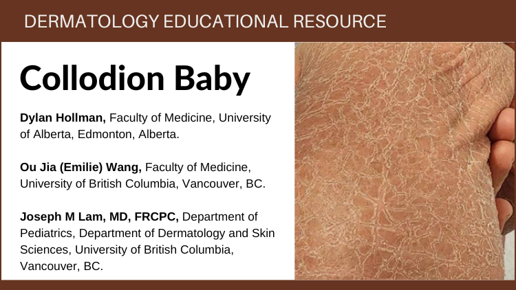
Collodion Baby

Teaser: 

Dylan Hollman,1Ou Jia (Emilie) Wang,2 Joseph M. Lam, MD, FRCPC,3

1Faculty of Medicine, University of Alberta.2 Faculty of Medicine, University of British Columbia, Vancouver, British Columbia, Canada.
3Department of Pediatrics, Department of Dermatology and Skin Sciences, University of British Columbia, Vancouver, British Columbia, Canada.

CLINICAL TOOLS

Abstract: Collodion baby, estimated to occur in 1 in 100,000 newborns, is a visually striking clinical presentation seen in neonates that is often a sign of an underlying autosomal recessive congenital ichthyosis. The baby is wrapped in a taut, translucent membrane, which is often compared to plastic wrap, saran wrap, a cocoon, or armour. A formal clinical diagnosis is often not reached until shedding of the membrane reveals the underlying phenotype. This can be isolated or associated with other structural and systemic congenital abnormalities. Patients may require ongoing monitoring and sometimes surgical intervention. Collodion baby is a rare and challenging condition that requires multimodal management including dermatologic care, infection prevention, nutritional support, developmental monitoring, and procedural interventions, if needed.
Key Words: Collodion baby, ichthyosis, neonate, newborn, pediatrics, dermatology.

Members of the College of Family Physicians of Canada may claim MAINPRO-M2 Credits for this unaccredited educational program.

www.cfpc.ca/Mainpro_M2

You can take quizzes without subscribing; however, your results will not be stored. Subscribers will have access to their quiz results for future reference.

Collodion baby is both a diagnosis and a clinical manifestation in newborns who commonly have autosomal recessive congenital ichthyosis.
A highly compromised skin barrier puts the patient at a high risk of both hypo-/hyperthermia, dehydration, poor growth, infection and several other organ-specific complications. Due to these increased risks, admission to the neonatal intensive care unit is necessary to facilitate close monitoring and access to a highly humidified incubator.
The collodion membrane (CM) is shed within 3 to 4 weeks, revealing the underlying ichthyosis. Special investigations can be undertaken before the membrane sheds such as a skin biopsy or blood work. These investigations can provide clinical clues to an earlier diagnosis. If the patient is stable, it is reasonable to wait for the membrane shedding to reveal an underlying diagnosis.
Petroleum-based moisturizers can protect the skin as the membrane peels off.
The most common underlying diagnoses of collodion baby are congenital ichthyosiform erythroderma and lamellar ichthyosis. However, an estimated 10% of patients will have near normal-appearing skin, referred to as self-improving collodion ichthyosis.
Skin barrier dysfunction can lead to significantly higher transepidermal water loss and poor temperature regulation. A highly humidified incubator (minimum 60%) can help reduce water loss and assist in adequate temperature regulation.
Other keys to management include close observation for signs of infection, dehydration, electrolyte imbalance and/or poor feeding/decreased growth velocity.
Topical petroleum-based lubricants should be applied multiple times per day while medicated ointments should be avoided due to risk of systemic toxicity.
Complications involving the lungs (chest constriction or respiratory distress), eyes (ectropion or keratitis) and ears (obstruction, conductive and sensorineural hearing loss) may also be seen. In these instances, consultations with pulmonology, ophthalmology or otorhinolaryngology may be necessary for comprehensive care.
Skin biopsy prior to membrane shedding is generally unhelpful but may provide disease-specific histological findings if done after the collodion sheds.
To have access to full article that these tools were developed for, please subscribe. The cost to subscribe is $80 USD per year and you will gain full access to all the premium content on www.healthplexus.net, an educational portal, that hosts 1000s of clinical reviews, case studies, educational visual aids and more as well as within the mobile app.
Disclaimer: 
Disclaimer at the end of each page

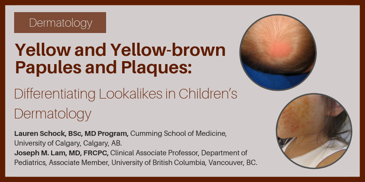
Yellow and yellow-brown papules and plaques: Differentiating look-alikes in children’s dermatology

Teaser: 

Lauren Schock, BSc, MD Program,1 Joseph M. Lam, MD, FRCPC,2

1Cumming School of Medicine, University of Calgary, Calgary, AB.
2Clinical Associate Professor, Department of Pediatrics, Associate Member, Department of Dermatology and Skin Sciences, University of British Columbia, Vancouver, BC.

CLINICAL TOOLS

Abstract: Yellow-hued papules and plaques in children can be difficult to differentiate as many causes are rare and may not be frequently outside of specialty pediatric dermatology settings. We will review some of the common and concerning yellow-brown papules and plaques found in infants and children and discuss appearance and distribution, pathophysiology, associated findings, and management.
Key Words: dermatology, pediatric, yellow lesions.
Nevus sebaceous typically grow in proportion with patients in early childhood. Excision should be deferred until adolescence to avoid the use of general anesthetic and an informed decision can be made by the child.
Benign cephalic histiocytosis and juvenile xanthogranuloma are both forms of non-Langerhans cell histiocytosis and are benign and self limited.
Consider a diagnosis of tuberous sclerosis in any child presenting with connective tissue nevi, especially if white macules, angiofibroma, or periungual fibroma are also found.
Screen children with necrobiosis lipodica for retinopathy and neuropathy.
Use your hands – rub a suspected lesion of mastocytosis; if urticaria is elicited (a red, itchy, swollen papule or plaque), you have found Darier's sign. Mastocytosis is likely. Be prepared to treat the child with antihistamines if needed.
Juvenile xanthogranulomas are more common under two years of age, and typically appear on the head and neck. Cutaneous xanthomas often occur overlying tendons, or as grouped papules over the extensor surfaces and buttocks.
To have access to full article that these tools were developed for, please subscribe. The cost to subscribe is $80 USD per year and you will gain full access to all the premium content on www.healthplexus.net, an educational portal, that hosts 1000s of clinical reviews, case studies, educational visual aids and more as well as within the mobile app.
Disclaimer: 
Disclaimer at the end of each page

About Psoriasis

About Psoriasis

WHAT IS PSORIASIS?

Psoriasis is a common but chronic skin condition that causes inflammation and scaling (red elevated patches and flaking silvery scales). The patches can be itchy or sore, causing discomfort and pain. Psoriasis causes skin cells to rise to the surface and shed at a very rapid rate. On average, people with psoriasis shed their skin cells every 3 to 4 days, while people without the condition have a turnover rate of about every 30 days.1,2,3,4
About Rosacea

About Rosacea

WHAT IS ROSACEA?

Rosacea is a common, chronic skin condition that causes redness of the face. It often presents as a mild redness or blushing that, over time, lasts for longer durations and becomes more pronounced. Rosacea can also produce enlarged, visible blood vessels and small red bumps on the facial skin. Before diagnosis, it can be mistaken for acne, an allergic reaction, or other skin conditions.1,2,3

Frequently Asked Questions about Psoriasis

Frequently Asked Questions about Psoriasis

WHAT IS PSORIASIS?

Psoriasis is a common but chronic skin condition that causes inflammation and scaling (red elevated patches and flaking silvery scales). The patches can be itchy or sore, causing discomfort and pain. Psoriasis causes skin cells to rise to the surface and shed at a very rapid rate. On average, people with psoriasis shed their skin cells every 3 to 4 days, while people without the condition have a turnover rate of about every 30 days.1,2,3,4

A Scaly Periorbital Rash in a Preschool-aged Boy

A Scaly Periorbital Rash in a Preschool-aged Boy

Teaser: 

Jennifer Smitten, MD, FRCPC,1 Joseph M Lam, MD, FRCPC,2

1BC Children's Hospital, University of British Columbia, BC.
2Assistant Clinical Professor, Department of Paediatrics, Associate Member, Department of Dermatology, University of British Columbia, BC.

CLINICAL TOOLS

Abstract: A healthy 4-year-old boy presented with an 8-month history of a pruritic scaly eruption around his right eye associated with several small pearly papules on the face. A clinical diagnosis of an eczematous id reaction to molluscum contagiosum was made. While up to 40% of cases of molluscum contagiosum may have an associated eczematous dermatitis, these are often under-recognized or misdiagnosed.
Key Words: Pediatrics, Dermatology, Dermatitis, Molluscum, Eczema, Id reaction, Viral exanthem, Hypersensitivity.
Eczematous id reactions to molluscum contagiosum (MC) in children are common, occurring in up to 40% of cases of MC.
Id reactions to MC can be challenging to diagnose, as they may occur at sites distant from the MC lesions.
Id reactions can be caused by a variety of infectious and noninfectious dermatoses.
Asymptomatic id reactions do not require pharmacologic treatment and a watchful waiting approach is reasonable.
1. Id reactions can be caused by a variety of infectious and noninfectious dermatoses, including allergic contact dermatitis to nickel, scabies infestation, tinea infection and molluscum infection.
2. In a unilateral eczematous dermatitis, consider molluscum dermatitis, especially in a child with no personal or family history of atopy.
3. Treatment of symptomatic id reactions may help to reduce spread of MC via autoinoculation from scratching.
To have access to full article that these tools were developed for, please subscribe. The cost to subscribe is $80 USD per year and you will gain full access to all the premium content on www.healthplexus.net, an educational portal, that hosts 1000s of clinical reviews, case studies, educational visual aids and more as well as within the mobile app.

Skin Manifestations of Internal Disease in Older Adults

Skin Manifestations of Internal Disease in Older Adults

Teaser: 

William Lear, MD, FRCPC, FAAD, Dermatologist, Silver Falls Dermatology PC, Salem, OR, USA.
Jennifer Akeroyd, RN, PhD student, Oregon Health & Science University, Portland, OR, USA.

In this article, we discuss skin findings affecting older adults, with a focus on pruritus, flushing, dermatitis, and ulcers, and consider related internal diseases. Our goal is to make this information readily transferable to the clinical setting for the non-dermatologist.
Key words: dermatology, skin manifestations, older adults, pruritus, flushing, dermatitis, ulcers.