Advertisement

Advertisement

Articles

Evaluation and Treatment of Sacroiliac Joint Dysfunction in the Primary Care Setting: A Practical Review

Teaser: 

Sydney Rucker, BS, 1 Adrienne Kelly, MD, FRCSC,2 David W. Polly, MD,3Robert J. Ferdon, MS,4 Robert A. Ravinsky, MDCM, MPH, FRCSC,5

1Medical University of South Carolina College of Medicine, Charleston, SC.
2 Orthopaedic Surgeon, Sault Area Hospital, Assistant Professor, Northern Ontario School of Medicine, Sault Ste Marie, ON.

3 Department of Orthopaedic Surgery, University of Minnesota, Minneapolis, MN, USA.

4 Department of Orthopaedics and Physical Medicine, Medical University of South Carolina, Charleston, SC, University of South Carolina School of Medicine, Columbia, SC.

5 Department of Orthopaedics and Physical Medicine, Medical University of South Carolina, Charleston, SC.

CLINICAL TOOLS

Abstract: Mechanical dysfunction of the sacroiliac joint (SIJ) is an often overlooked, but a common cause of low back pain in the North American adult population. The diagnosis is primarily clinical and requires the exclusion of other potential etiologies of low back pain (LBP). A number of non-surgical treatment options are available for patients with this pathological entity. In cases of persistent, severe SIJ pain refractory to non-operative measures, SIJ fusion may be considered as a surgical intervention.
Key Words: sacroiliitis, sacroiliac dysfunction, sacroiliac joint (SIJ), low back pain (LBP), gluteal pain, SI joint fusion, percutaneous SI joint fixation.

Members of the College of Family Physicians of Canada may claim MAINPRO-M2 Credits for this unaccredited educational program.

www.cfpc.ca/Mainpro_M2

You can take quizzes without subscribing; however, your results will not be stored. Subscribers will have access to their quiz results for future reference.

1. The SI joint is a known contributor to low back pain
2. Non-surgical treatment remains the primary management approach for SIJ dysfunction
3. Diagnostic intra-articular injections performed under image guidance are considered the gold standard for confirming SIJ-mediated pain
4. For patients with clinically debilitating mechanical SIJ dysfunction who have failed an exhaustive course of non-operative treatment, surgical options may be considered
1. SIJ dysfunction is a known contributor to low back pain, accounting for approximately 15-30% of LBP cases in the outpatient setting.
2. While imaging plays a role in excluding alternative diagnoses, no imaging modality has demonstrated reliable diagnostic utility for mechanical SIJ dysfunction although there is some utility of MRI in the setting of inflammatory sacroiliitis.
3. Patients with SIJ pain typically report symptoms consistently localized to an area within 1cm inferomedial to the posterior superior iliac spine (PSIS) that may radiate into the buttocks, groin, posterior thigh or even past the knee and into the foot.
4. Diagnosis is by physical examination which should include a variety of SIJ-specific provocation tests. Three or more positive results out of five standardized maneuvers is supported by Level 1 evidence for a Clinical Diagnostic Rule.
To have access to full article that these tools were developed for, please subscribe. The cost to subscribe is $80 USD per year and you will gain full access to all the premium content on www.healthplexus.net, an educational portal, that hosts 1000s of clinical reviews, case studies, educational visual aids and more as well as within the mobile app.

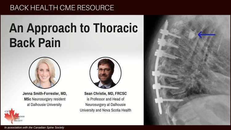
An Approach to Thoracic Back Pain

Teaser: 

Jenna Smith-Forrester, MD, MSc, 1 Sean Christie, MD, FRCSC,2

1Neurosurgery resident at Dalhousie University.
2 is Professor and Head of Neurosurgery at Dalhousie University and Nova Scotia Health.

CLINICAL TOOLS

Abstract: Thoracic back pain, while less prevalent than cervical or lumbar pain, encompasses a wide spectrum of conditions ranging from benign to life-threatening. This article discusses both non-spine-related causes, such as aortic dissection and pancreatitis, as well as spine-related pathology including compression fractures, herpetic neuralgia, thoracic disc herniations, spinal metastases, and intradural lesions like meningiomas and syringomyelia. Using clinical pearls, case studies, and evidence-based approaches, this article offers primary healthcare providers with the tools to diagnose and manage thoracic back pain effectively.
Key Words: Thoracic back pain, thoracic disc herniation, spinal metastasis, spinal compression fracture, syringomyelia (Syrinx).

Members of the College of Family Physicians of Canada may claim MAINPRO-M2 Credits for this unaccredited educational program.

www.cfpc.ca/Mainpro_M2

You can take quizzes without subscribing; however, your results will not be stored. Subscribers will have access to their quiz results for future reference.

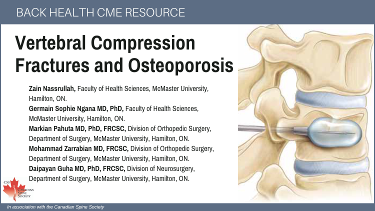
Spinal Compression Fractures: Most commonly caused by osteoporosis, compression fractures present with acute pain exacerbated by movement. Prevention efforts focus on frailty screening and osteoporosis management.
Herpetic Neuralgia: Thoracic dermatomes are frequently affected, with dermatomal pain and vesicular rash often confirming the diagnosis. Neuropathic pain agents remain the cornerstone of treatment.
Thoracic Disc Herniation: Though rare (1:1,000,000), can cause thoracic pain and radiculopathy and lead to myelopathy with spastic paraparesis. MRI is the gold standard for diagnosis, with surgical interventions considered for refractory cases.
Spinal Metastasis: The thoracic spine is the most common site for spinal metastases, presenting with nocturnal pain, neurologic deficits, and instability. Treatment involves palliation, pain relief, and spinal stabilization. 
Thoracic viscera may be the source of sudden severe pain, often associated with hemodynamic or constitutional symptoms.
MRI is the preferred imaging modality for diagnosing thoracic disc herniation, spinal cord compression, and intramedullary lesions.
Early recognition and referral to specialists are crucial for conditions like thoracic disc herniation, unstable spinal metastatic disease, and syrinx to improve patient outcomes.
To have access to full article that these tools were developed for, please subscribe. The cost to subscribe is $80 USD per year and you will gain full access to all the premium content on www.healthplexus.net, an educational portal, that hosts 1000s of clinical reviews, case studies, educational visual aids and more as well as within the mobile app.

Keratosis Pilaris

Teaser: 

Bianca Te, BSc,1Joseph M. Lam, MD, FRCPC,2

1Faculty of Medicine, University of British Columbia, Vancouver, BC, Canada.2 Department of Pediatrics, Department of Dermatology and Skin Sciences, University of British Columbia, Vancouver, British Columbia, Canada.

CLINICAL TOOLS

Abstract: Keratosis pilaris (KP) is a common, benign skin condition that primarily affects children and adolescents. It is characterized by small follicular papules resembling “chicken skin” or goosebumps, most often on extensor surfaces of the proximal upper and lower extremities. The exact cause of KP is unclear, however it is believed to follow an autosomal dominant inheritance pattern and may be associated with a mutation in filaggrin, a protein involved in maintaining the skin barrier. This ultimately results in excessive keratin production and deposition surrounding hair follicles. Diagnosis is typically based on the patient’s history and clinical presentation. KP is most often asymptomatic and tends to improve with age. However, treatment is aimed at enhancing cosmetic appearance or relieving mild pruritus. Management includes optimizing skin moisturization and using topical keratolytic agents, though other options such as topical retinoids and laser therapy may also be effective.
Key Words: Keratosis pilaris, keratolytic agents, asymptomatic, benign.

Members of the College of Family Physicians of Canada may claim MAINPRO-M2 Credits for this unaccredited educational program.

www.cfpc.ca/Mainpro_M2

You can take quizzes without subscribing; however, your results will not be stored. Subscribers will have access to their quiz results for future reference.

Keratosis pilaris presents as numerous small follicular papules resembling “chicken skin” or goosebumps, most often on extensor surfaces of the proximal upper and lower extremities.
Keratosis pilaris is a benign skin condition that often improves with age.
Management of keratosis pilaris is aimed at reducing symptoms such as pruritus and improving cosmetic appearance. Keratolytic agents such as lactic acid or salicylic acid creams are most commonly used for treatment.
Keratosis pilaris is the result of excess keratin production that deposits around the hair follicle, however the exact pathogenesis is not fully understood.
Diagnosis of keratosis pilaris is made on history and physical exam. Dermoscopy and biopsy are not often required, but can be used if other conditions are suspected.
Patients should understand that keratosis pilaris is benign and that there is no cure. It is important to provide reassurance that the condition often improves with age, however some topical treatments may be able to help improve cosmetic appearance.
To have access to full article that these tools were developed for, please subscribe. The cost to subscribe is $80 USD per year and you will gain full access to all the premium content on www.healthplexus.net, an educational portal, that hosts 1000s of clinical reviews, case studies, educational visual aids and more as well as within the mobile app.
Disclaimer: 
Disclaimer at the end of each page

An Approach to the Differential Diagnosis of Sciatica in the Primary Care Setting

Teaser: 

Taryn Walker B.Kin, MM, 1 Vahagn Karapetyan MD PhD FRCSC,2 Greg McIntosh MSc,3 Christopher S Bailey MD FRCSC,4

1Department of Surgery, Schulich School of Medicine, Western University
2Department of Surgery, Schulich School of Medicine, Western University
3Director of Research Operations, Canadian Spine Outcomes and Research Network
4Department of Surgery, Schulich School of Medicine, Western University

CLINICAL TOOLS

Abstract: Sciatica is a well-recognized complaint, with the hallmark presenting symptom of lancinating pain running down the back of the leg, along the path of the sciatic nerve. While it is typically caused by a herniated lumbar disc within the spinal canal, an understanding of the course of the sciatic nerve and associated regional anatomy is useful in forming a broader differential diagnosis. In rare cases, sciatica, usually bilateral, is associated with a loss of bowel or bladder function, indicating cauda equina syndrome, a medical emergency. Other degenerative conditions of the lumbar spine such as spondylolisthesis and spinal stenosis can occasionally produce sciatica while deep gluteal pain is often used to describe the muscular causes. Malignancy, trauma, vascular causes and ectopic endometrial tissue can cause compression of the lumbosacral plexus within the pelvis and produce similar symptoms. In this review, we highlight the common clinical presentations, physical examination and relevant diagnostic investigations for a broad differential diagnosis of sciatica.
Key Words: Sciatica, leg dominant pain, nerve compression, straight leg raise.

Members of the College of Family Physicians of Canada may claim MAINPRO-M2 Credits for this unaccredited educational program.

www.cfpc.ca/Mainpro_M2

You can take quizzes without subscribing; however, your results will not be stored. Subscribers will have access to their quiz results for future reference.

1. Herniated intervertebral disc accounts for up to 90% of sciatica cases 
2. An understanding of the course of the sciatic nerve and associated regional anatomy can be useful in forming a differential diagnosis; one should consider gluteal region sciatic compression disorders, pelvic pathology involving the lumbosacral plexus, malignancy, infection, trauma and conditions that mimic sciatica.
3. Patients usually have low back pain associated with their leg complaints, but genuine sciatica from a herniated lumbar disc produces leg pain that is the dominant symptom. Back dominant pain, with any direct nerve involvement, can extend into the leg, occasionally all the way to the foot, but unless the leg pain is more intense than the back pain, it is not sciatica. 
4. The physical examination should include checking lower limb reflexes, dermatome and myotome assessment, upper motor neuron tests such as the plantar response and specialized neural tension tests such as the passive straight leg raise (SLR) and slump tests.
5. A positive crossover sign occurs when raising the straight leg on the affected side reproduces not only the typical pain in the affected leg but causes pain to radiate down the seemingly unaffected leg as well. This finding of bilateral sciatica suggests pathology located in the midline, a central disc herniation.
True sciatica from a herniated lumbar disc is when the leg pain is the dominant symptom over back pain. Lancinating, constant, radicular pain down the posterior leg along the path of the sciatic nerve is the hallmark symptom. Activity, prolonged sitting or Valsalva maneuvers such as coughing or bearing down aggravate sciatic pain. A patient may prefer to stand because sitting is intolerable.
Neurogenic Claudication from spinal stenosis is distinctive from herniated lumbar disc sciatica. Unlike classic sciatica, lumbar spinal stenosis neural tension tests are often negative and the neurological assessment may be normal; however, the more classic sciatic presentation, may be present in spinal stenosis if a focal nerve root is also compressed.
To distinguish piriformis syndrome from sciatica, muscle tenderness with deep gluteal palpation and specialized piriformis stretch tests such as the FAIR, Pace and Beatty maneuvers may be helpful.
Greater trochanteric bursitis, meralgia paresthetica, and cluneal nerve entrapment are conditions that can cause pain in a distribution that is similar to the sciatic nerve, but without involving the nerve directly.
Identifying any posterior thigh pain when the leg is elevated above 60 or 70 degrees as sciatica is a common diagnostic error. Unless the leg pain produced is identical to the patient’s chief complaint on history, the test is negative.
To have access to full article that these tools were developed for, please subscribe. The cost to subscribe is $80 USD per year and you will gain full access to all the premium content on www.healthplexus.net, an educational portal, that hosts 1000s of clinical reviews, case studies, educational visual aids and more as well as within the mobile app.

Understanding Myofascial Pain Syndrome: Causes, Diagnosis, and Treatment

Teaser: 

Eugene K. Wai MD, MSc, CIP, FRCSC, 1 Ted Findlay, DO, CCFP, FCFP,2

1Associate Professor, Division of Orthopaedic Surgery, Cross Appointment to School of Epidemiology and Public Health, University of Ottawa, Ottawa, ON.
2Clinical Associate Professor, Program Director, Family Medicine Chronic Pain Enhanced Skills R3, Department of Family Medicine, University of Calgary, Calgary, AB.

CLINICAL TOOLS

Abstract: Myofascial Pain Syndrome (MPS) is a chronic pain disorder characterized by localized pain originating from myofascial trigger points (MTrPs) within the skeletal muscle of the spine and should be included in the differential diagnosis for non-surgical back pain. The etiology of MPS is multifactorial, involving trauma, repetitive strain, and postural dysfunction, leading to the formation of hyperirritable nodules that cause both local and referred pain. Diagnosis is primarily clinical, relying on the identification of MTrPs through physical examination. Treatment approaches include pharmacological interventions, manual therapies, and needling techniques. Evidence for long-term efficacy remains limited. Future research is essential to establish reliable diagnostic criteria and effective treatment modalities for MPS.
Key Words: Myofascial pain syndrome, trigger points, diagnosis, treatment, chronic pain, musculoskeletal disorders, manual therapy, pharmacological interventions.

Members of the College of Family Physicians of Canada may claim MAINPRO-M2 Credits for this unaccredited educational program.

www.cfpc.ca/Mainpro_M2

You can take quizzes without subscribing; however, your results will not be stored. Subscribers will have access to their quiz results for future reference.

1. Myofascial Pain Syndrome (MPS) is characterized by the presence of hyperirritable nodules, or myofascial trigger points, within taut bands of skeletal muscle, leading to localized and referred pain.
2. MPS can be caused by factors such as trauma, repetitive strain, poor posture, and muscle deconditioning, with theories suggesting muscle fiber energy crises or neurogenic inflammation as the main contributors.
3. The diagnosis of MPS is primarily clinical, relying on physical examination including palpable nodules, local twitch responses, and reproduction of the patient’s pain with trigger point palpation. There are no standard imaging or lab tests.
4. Treatment includes a combination of exercise, manual therapy, pharmacological interventions, trigger point injections and dry needling. Dry needling has shown the most promise.
5. There is no established Clinical Practice Guideline for MPS, and high-quality evidence supporting the long- term efficacy of current treatments is lacking. Further research into pathophysiology and treatment strategies is needed.
1. Trigger points are active or latent—active points cause spontaneous pain and limit muscle function, while latent points are asymptomatic until palpated.
2. MPS involves localized pain and trigger points; fibromyalgia presents with widespread pain and central sensitization. Conditions may coexist but require different management strategies.
3. A multimodal approach, combining dry needling with other physical therapies, yields better outcomes compared to single-modality treatments.
4. Opioids have limited evidence of efficacy and the potential to delay recovery. Use non-opioid and non-invasive interventions.
To have access to full article that these tools were developed for, please subscribe. The cost to subscribe is $80 USD per year and you will gain full access to all the premium content on www.healthplexus.net, an educational portal, that hosts 1000s of clinical reviews, case studies, educational visual aids and more as well as within the mobile app.

Symptomatic Lumbar Canal Stenosis—A Review and Primer on Surgical Decision Making

Teaser: 

Sager Hanna MB, BCh, BAO, 1 Perry Dhaliwal MD, MPH, FRCSC,2

1Section of Neurosurgery and Section of Orthopedic Surgery, Department of Surgery, Rady Faculty of Health Sciences, University of Manitoba.
2Assistant Professor of Neurosurgery, Section of Neurosurgery, Department of Surgery, Rady Faculty of Health Sciences, University of Manitoba.

CLINICAL TOOLS

Abstract: Lumbar canal stenosis is an anatomical term used to describe narrowing of the spinal canal either congenitally or from age-related degenerative changes. It refers to a structural finding that may or may not be symptomatic. A decrease in canal diameter can lead to compression of the neural components, causing a constellation of symptoms. Family physicians should familiarize themselves with the various presentations of canal narrowing and the available diagnostic and treatment options.
Key Words: lumbar spinal stenosis, neurogenic claudication, back pain, radiculopathy.

Members of the College of Family Physicians of Canada may claim MAINPRO-M2 Credits for this unaccredited educational program.

www.cfpc.ca/Mainpro_M2

You can take quizzes without subscribing; however, your results will not be stored. Subscribers will have access to their quiz results for future reference.

1. Lumbar spinal stenosis is commonly caused by age-related degenerative changes involving the intervertebral discs, ligamentum flavum and facet joints.
2. Patients with lumbar spinal stenosis may present with neurogenic claudication or radiculopathy.
3. The primary care provider needs to distinguish between symptomatic lumbar spinal stenosis and other common mimics.
4. Surgical treatment is principally decompression of the neural elements with the possible addition of fusion of the affected levels.
1. Degenerative changes in the lumbar spine can lead to various symptoms such as low back pain, lumbar radiculopathy, neurogenic claudication, and cauda equina syndrome.
2. Imaging of the lumbar spine should be ordered when there is a high clinical suspicion of lumbar spinal canal stenosis based on the history and physical examination.
3. Initial management of patients presenting with lumbar canal stenosis involves non-operative modalities like pharmacological therapy, physiotherapy, lifestyle modifications, patient education and image-guided injections.
4. Surgical decompression for symptomatic lumbar spinal stenosis, with or without fusion, is generally indicated when symptoms significantly interfere with daily activity and non-operative treatment has failed after 3-6 months.
To have access to full article that these tools were developed for, please subscribe. The cost to subscribe is $80 USD per year and you will gain full access to all the premium content on www.healthplexus.net, an educational portal, that hosts 1000s of clinical reviews, case studies, educational visual aids and more as well as within the mobile app.

Collodion Baby

Teaser: 

Dylan Hollman,1Ou Jia (Emilie) Wang,2 Joseph M. Lam, MD, FRCPC,3

1Faculty of Medicine, University of Alberta.2 Faculty of Medicine, University of British Columbia, Vancouver, British Columbia, Canada.
3Department of Pediatrics, Department of Dermatology and Skin Sciences, University of British Columbia, Vancouver, British Columbia, Canada.

CLINICAL TOOLS

Abstract: Collodion baby, estimated to occur in 1 in 100,000 newborns, is a visually striking clinical presentation seen in neonates that is often a sign of an underlying autosomal recessive congenital ichthyosis. The baby is wrapped in a taut, translucent membrane, which is often compared to plastic wrap, saran wrap, a cocoon, or armour. A formal clinical diagnosis is often not reached until shedding of the membrane reveals the underlying phenotype. This can be isolated or associated with other structural and systemic congenital abnormalities. Patients may require ongoing monitoring and sometimes surgical intervention. Collodion baby is a rare and challenging condition that requires multimodal management including dermatologic care, infection prevention, nutritional support, developmental monitoring, and procedural interventions, if needed.
Key Words: Collodion baby, ichthyosis, neonate, newborn, pediatrics, dermatology.

Members of the College of Family Physicians of Canada may claim MAINPRO-M2 Credits for this unaccredited educational program.

www.cfpc.ca/Mainpro_M2

You can take quizzes without subscribing; however, your results will not be stored. Subscribers will have access to their quiz results for future reference.

Collodion baby is both a diagnosis and a clinical manifestation in newborns who commonly have autosomal recessive congenital ichthyosis.
A highly compromised skin barrier puts the patient at a high risk of both hypo-/hyperthermia, dehydration, poor growth, infection and several other organ-specific complications. Due to these increased risks, admission to the neonatal intensive care unit is necessary to facilitate close monitoring and access to a highly humidified incubator.
The collodion membrane (CM) is shed within 3 to 4 weeks, revealing the underlying ichthyosis. Special investigations can be undertaken before the membrane sheds such as a skin biopsy or blood work. These investigations can provide clinical clues to an earlier diagnosis. If the patient is stable, it is reasonable to wait for the membrane shedding to reveal an underlying diagnosis.
Petroleum-based moisturizers can protect the skin as the membrane peels off.
The most common underlying diagnoses of collodion baby are congenital ichthyosiform erythroderma and lamellar ichthyosis. However, an estimated 10% of patients will have near normal-appearing skin, referred to as self-improving collodion ichthyosis.
Skin barrier dysfunction can lead to significantly higher transepidermal water loss and poor temperature regulation. A highly humidified incubator (minimum 60%) can help reduce water loss and assist in adequate temperature regulation.
Other keys to management include close observation for signs of infection, dehydration, electrolyte imbalance and/or poor feeding/decreased growth velocity.
Topical petroleum-based lubricants should be applied multiple times per day while medicated ointments should be avoided due to risk of systemic toxicity.
Complications involving the lungs (chest constriction or respiratory distress), eyes (ectropion or keratitis) and ears (obstruction, conductive and sensorineural hearing loss) may also be seen. In these instances, consultations with pulmonology, ophthalmology or otorhinolaryngology may be necessary for comprehensive care.
Skin biopsy prior to membrane shedding is generally unhelpful but may provide disease-specific histological findings if done after the collodion sheds.
To have access to full article that these tools were developed for, please subscribe. The cost to subscribe is $80 USD per year and you will gain full access to all the premium content on www.healthplexus.net, an educational portal, that hosts 1000s of clinical reviews, case studies, educational visual aids and more as well as within the mobile app.
Disclaimer: 
Disclaimer at the end of each page

Scoliosis Screening: A Review of Current Evidence, Worldwide Practices, and Recommendations for Implementation Across Canada

Teaser: 

Caitlyn Dunphy, MPT, 1 Marie Anne Keenan, BSc candidate,2 Hunter Cole David Arulpragasam, BSc candidate,3 Jean Albert Ouellet, MD, FRCS(C),4 Kevin Smit, MD, FRCS(C),5 Ron El-Hawary, MD, MSc, FRCS(C),6 Andrea Mary Simmonds, MD, MHSc, FRCS(C),7

1BC Children’s Hospital Orthopaedic Spine Clinic.
2University of Victoria, Research Student, Department of Orthopaedic Surgery, BC Children’s Hospital.
3University of Toronto, Research Student, Department of Orthopaedic Surgery, BC Children’s Hospital.
4McGill University Health Centre/ Shriners Hospital for Children - Canada.
5Pediatric Orthopedic Surgeon, CHEO, Associate Professor, Faculty of Medicine, University of Ottawa, Surgeon Scientist, CHEO Research Institute.
6Professor of Surgery (Orthopedics, Neurosurgery) Professor of Biomedical Engineering, Faculty of Medicine, Dalhousie University Chief of Pediatric Orthopedic Surgery, IWK Health.
7 Paediatric Spine & Orthopaedic Trauma Surgeon, British Columbia Children’s Hospital Clinical Assistant Professor, UBC Department of Orthopaedics.

CLINICAL TOOLS

Abstract: There is a lack of consensus about the merits of scoliosis screening and whether it is a beneficial strategy for both the patients and the healthcare system. With mounting concerns about long wait times across Canada for surgical correction of scoliosis, interest has grown in maximizing non-operative care. We have investigated the history of scoliosis screening and the controversies surrounding implementation of screening in a Canadian setting. We propose an optimal screening strategy.
Key Words: Scoliosis, scoliosis screening, early detection, conservative strategies.

Members of the College of Family Physicians of Canada may claim MAINPRO-M2 Credits for this unaccredited educational program.

www.cfpc.ca/Mainpro_M2

You can take quizzes without subscribing; however, your results will not be stored. Subscribers will have access to their quiz results for future reference.

Screening can facilitate early diagnosis and treatment of scoliosis.
Early diagnosis of scoliosis increases opportunities for successful conservative treatment.
Conservative strategies may prevent the need for surgical intervention.
Scoliosis screening may improve access to care and reduce health care costs.
Early detection of scoliosis through school screenings is recommended for initiating timely and effective conservative treatments, such as bracing and physical therapy. This can significantly reduce the need for surgical interventions and associated healthcare costs.
A standardized, evidence-based screening protocol should be developed and implemented across all Canadian schools. This protocol should include clear guidelines on the use of screening tools, referral criteria, and follow-up procedures to ensure consistency and accuracy in detecting scoliosis.
School nurses, physical education teachers, and other relevant personnel should receive adequate resources and support for proper training in scoliosis screening.
Educational campaigns must raise awareness about the signs of scoliosis and the importance of school screenings for early detection among parents, teachers, and the general public.
Ongoing research and evaluation of the screening program should be conducted to assess its effectiveness, cost-benefit ratio, and impact on health outcomes.
Collaboration between healthcare providers, educators, policymakers, and scoliosis advocacy groups is essential to create a comprehensive and sustainable screening program.
To have access to full article that these tools were developed for, please subscribe. The cost to subscribe is $80 USD per year and you will gain full access to all the premium content on www.healthplexus.net, an educational portal, that hosts 1000s of clinical reviews, case studies, educational visual aids and more as well as within the mobile app.

Pityriasis Alba

Teaser: 

Ou Jia (Emilie) Wang,1 Joseph M. Lam, MD, FRCPC,2

1 Faculty of Medicine, University of British Columbia, Vancouver, British Columbia, Canada.
2Department of Pediatrics, Department of Dermatology and Skin Sciences, University of British Columbia, Vancouver, British Columbia, Canada.

CLINICAL TOOLS

Abstract: Pityriasis alba is a common, benign skin condition that primarily affects children and adolescents, characterized by hypopigmented patches and scaly plaques on the face and other areas of the body. It is likely a manifestation of post-inflammatory hypopigmentation from subtle or subclinical inflammation. Diagnosis is typically based on history and clinical presentation. Management involves the use of emollients and low-potency topical steroids to improve skin hydration, reduce inflammation, and alleviate symptoms such as pruritus. Pityriasis alba typically becomes less apparent as the patients age, but reassurance and symptomatic relief are critical components to managing the condition.
Key Words: Pityriasis alba, atopy, hypersensitivity, scaling, hypopigmentation, asymptomatic.

Members of the College of Family Physicians of Canada may claim MAINPRO-M2 Credits for this unaccredited educational program.

www.cfpc.ca/Mainpro_M2

You can take quizzes without subscribing; however, your results will not be stored. Subscribers will have access to their quiz results for future reference.

Pityriasis alba presents as patches and plaques of hypopigmentation, which is more identifiable in darker skin types (Fitzpatrick skin types III to VI).
Pityriasis alba is a benign and self-limiting skin condition that often improves with time.
Pityriasis alba is often associated with atopic dermatitis and the atopic triad.
Diagnosis of pityriasis alba is made on history and exam and the exclusion of other conditions (e.g. fungal infections, atopic dermatitis, and psoriasis). Skin biopsy, laboratory tests, and Wood’s lamp examination are not necessary, but can be performed if other conditions are suspected.
The hypopigmentation in pityriasis alba does not result from reduction in melanocyte count.
Patient reassurance, education and lifestyle management is often sufficient, but emollients, low-potency topical steroids, and topical calcineurin can also be used.
To have access to full article that these tools were developed for, please subscribe. The cost to subscribe is $80 USD per year and you will gain full access to all the premium content on www.healthplexus.net, an educational portal, that hosts 1000s of clinical reviews, case studies, educational visual aids and more as well as within the mobile app.
Disclaimer: 
Disclaimer at the end of each page

Back and Neck Pain, Pain Clinics and Interventional Pain Management in Canada

Teaser: 

Arani Kulamurugan,1 Pranjan Gandhi,2 Markian Pahuta,3 Mohammad Zarrabian,4 Daipayan Guha,5

1Michael DeGroote School of Medicine, McMaster University, Hamilton, ON.
2Michael DeGroote School of Medicine, McMaster University, Hamilton, ON.
3Department of Surgery, Division of Orthopaedics, McMaster University, Hamilton, ON.
4Department of Surgery, Division of Orthopaedics, McMaster University, Hamilton, ON.
5Department of Surgery, Division of Neurosurgery, McMaster University, Hamilton, ON.

CLINICAL TOOLS

Abstract: This paper examines the role of pain clinics in Canada, focusing on non-surgical interventions to manage cervical and lumbar degenerative pathologies. These pathologies have a substantial impact on health care and the economy. Since non-interventional management strategies are often insufficient, pain clinics can be effective in providing image-guided injections to reduce symptoms and rates of surgery. Given the challenges of access and long wait times for treatment, the expansion of pain clinics may be an interim solution to improve outcomes and alleviate the burden on Canadian healthcare.
Key Words: radiculopathy, myelopathy, back pain, neck pain, pain clinic.

Members of the College of Family Physicians of Canada may claim MAINPRO-M2 Credits for this unaccredited educational program.

www.cfpc.ca/Mainpro_M2

You can take quizzes without subscribing; however, your results will not be stored. Subscribers will have access to their quiz results for future reference.

1. Identifying the specific type of back pain guides the choice of treatment, enhancing patient outcomes.
2. Interventional strategies have demonstrated significant benefits when combined with traditional medical and physical therapies.
3. Axial pain, radiculopathy, neurogenic claudication and myelopathy have distinct symptoms and relief mechanisms, making accurate diagnosis critical.
4. Improving the distribution and accessibility of multidisciplinary pain management services will improve the outcomes for patients with chronic pain.
Differentiating Pain Syndromes: It is essential to distinguish among axial neck/back pain, radicular pain, neurogenic claudication and myelopathy to institute proper back pain management. Axial pain is worsened by physical activity, radicular pain is limb dominant, neurogenic claudication is exacerbated by prolonged standing and relieved by sitting, and myelopathy produces upper motor neuron findings in both upper and lower limbs.
Role of Pain Clinics: Offering a wide range of services, pain clinics are cost-effective and improve quality of life and functionality through interventional pain management, mental health support, and physical therapy.
Barriers to Accessing Care: Access to multidisciplinary pain treatment facilities in Canada is limited by long wait times and significant regional variability.
To have access to full article that these tools were developed for, please subscribe. The cost to subscribe is $80 USD per year and you will gain full access to all the premium content on www.healthplexus.net, an educational portal, that hosts 1000s of clinical reviews, case studies, educational visual aids and more as well as within the mobile app.