Advertisement

Advertisement

kyphoplasty

Percutaneous Vertebral Augmentation for the Treatment of Pathological Fractures of the Spine

Teaser: 

Ayoub Dakson, MBChB, MSc, FRCSC,1 Sean Christie, MD, FRCSC,2

1Clinical Fellow, Department of Surgery (Neurosurgery) QEII Health Sciences Centre, Halifax, Nova Scotia.2Professor, Department of Surgery (Neurosurgery), QEII Health Sciences Centre, Halifax, Nova Scotia.

CLINICAL TOOLS

Abstract:Pathological vertebral fractures (PVFs) occur commonly due to osteoporosis or a metastatic lesion to the spine, and present with acute back pain and loss of independent ambulation. Appropriate clinical assessment and radiographic evaluation are required to ensure optimal patient selection for a percutaneous vertebral augmentation procedure (PVA). This review explores the pathogenesis of PVFs and the efficacy of PVA in improving pain-related outcomes as well as health-related quality of life scores in both osteoporotic and metastatic PVFs.
Key Words: Osteoporosis; pathological vertebral fractures; vertebroplasty; kyphoplasty.

Members of the College of Family Physicians of Canada may claim MAINPRO-M2 Credits for this unaccredited educational program.

www.cfpc.ca/Mainpro_M2

You can take quizzes without subscribing; however, your results will not be stored. Subscribers will have access to their quiz results for future reference.

Pathological vertebral fractures occur commonly due to osteoporosis and metastatic disease to the spine.
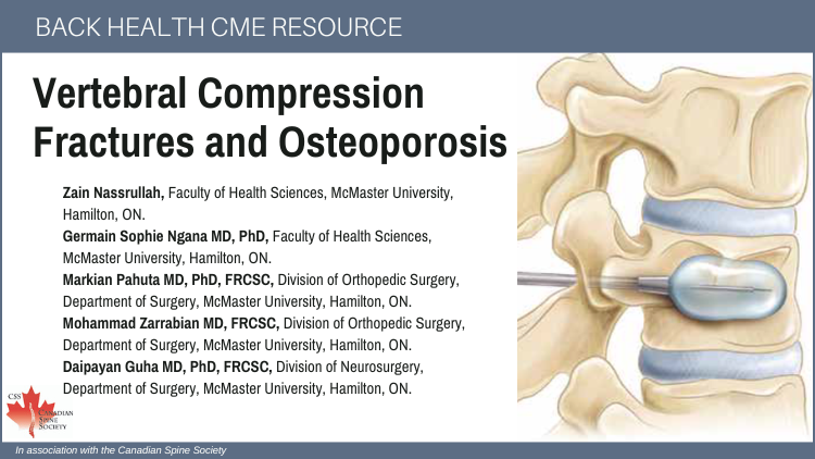
Percutaneous vertebral augmentation procedures consist of vertebroplasty or balloon-kyphoplasty with the goals of increasing the strength of fractured vertebral body and restoring its height in order to alleviate back pain and increase ambulation.
Balloon-kyphoplasty has been shown to improve back pain associated with PVFs and health-related quality of life scores.
Appropriate consideration of ''red flag'' features in the clinical history and neurologic examination of a patient with back pain is crucial in screening for a potential sinister underlying etiology (i.e. malignant pathological vertebral fractures with spinal cord compression, infection, etc.).
MRI imaging (STIR) may provide useful information in deciding if the fracture has already healed.
Loss of the integrity of the dorsal wall of the fractured vertebral body increases the risk of leakage of the injected cement into the spinal canal, potentially causing spinal cord compression.
To have access to full article that these tools were developed for, please subscribe. The cost to subscribe is $80 USD per year and you will gain full access to all the premium content on www.healthplexus.net, an educational portal, that hosts 1000s of clinical reviews, case studies, educational visual aids and more as well as within the mobile app.
Disclaimer: 
Disclaimer at the end of each page

Osteoporotic Vertebral Compression Fractures: Diagnosis and Management

Osteoporotic Vertebral Compression Fractures: Diagnosis and Management

Teaser: 

Michael M.H. Yang, MD, M.Biotech,1 W. Bradley Jacobs, MD, FRCSC,2

1Division of Neurosurgery, Department of Clinical Neuroscience, University of Calgary, Calgary, Alberta, Canada.
2Division of Neurosurgery, Department of Clinical Neuroscience, University of Calgary, Calgary, Alberta, Canada.

CLINICAL TOOLS

Abstract: Osteoporotic vertebral compression fractures (VCFs) are the most common fragility fracture and have significant impact on numerous indices of health quality. High risks patients should be identified and appropriate preventative therapy initiated. The majority of VCFs can be managed in a non-operative fashion, with analgesia as required to support progressive mobilization. Patients who fail non-operative measures may be considered for percutaneous vertebral augmentation. However, the efficacy of these procedures in altering the natural history of recovery is controversial. Surgery has a limited role in the initial management of VCFs and is typically restricted to the rare circumstance of VCF associated with acute neurological dysfunction.
Key Words: osteoporosis, vertebral compression fracture, vertebroplasty, kyphoplasty.

Members of the College of Family Physicians of Canada may claim MAINPRO-M2 Credits for this unaccredited educational program.

www.cfpc.ca/Mainpro_M2

You can take quizzes without subscribing; however, your results will not be stored. Subscribers will have access to their quiz results for future reference.

1. Osteoporosis is under diagnosed in Canada. Early diagnosis, fragility fracture risk stratification and initiation of preventative treatment is important, as osteoporotic vertebral compression fractures (VCFs) have a significant associated personal and societal health utility cost.
2. Patients suspected of having a VCF should have an AP and lateral X-ray of the suspected region. If VCF is confirmed, an upright X-ray should be performed to assess for stability. CT and/or MR imaging has limited utility in the absence of red flag signs or symptoms.
3. VCFs should be managed with initiation of an appropriate pain management regiment, early bed rest as required for pain control and gradual mobilization. Patients with refractory pain 4–6 weeks after onset can be considered for percutaneous vertebral cement augmentation (e.g. vertebroplasty), although the clinical efficacy of such procedures remains unclear.
A few screening measurements can be performed in the office setting to help significantly improve the likelihood of detecting a VCF on radiological studies. They include prospective height loss of greater than 2cm or a height loss, or a height loss based on history of more than 6cm, a rib-to-pelvis distance of less than 2 fingerbreadths, or an occipital-to-wall distance greater than 5cm.
Most patients with osteoporotic VCFs do not need a referral to a spine surgeon. Acute pain from a new VCF usually improves over a period of 6 weeks. Non-operative management should follow the WHO analgesic ladder starting with acetaminophen/NSAIDs followed by opioids, as necessary. The goal of treatment is to provide pain relief and facilitate early functional rehabilitation.
Patients with high or medium 10-year fracture risk should be considered for pharmacotherapy to prevent the progression of low bone mineral density and osteoporotic fractures.
To have access to full article that these tools were developed for, please subscribe. The cost to subscribe is $80 USD per year and you will gain full access to all the premium content on www.healthplexus.net, an educational portal, that hosts 1000s of clinical reviews, case studies, educational visual aids and more as well as within the mobile app.

Kyphoplasty and Vertebroplasty for the Treatment of Osteoporotic Vertebral Compression Fractures

Kyphoplasty and Vertebroplasty for the Treatment of Osteoporotic Vertebral Compression Fractures

Teaser: 

Karen Beattie, BSc, PhD Candidate and Dr. A. Papaioannou, MSc, MD, FRCP(C), Associate Professor of Medicine; Department of Geriatrics, McMaster University, Hamilton, ON.
Dr. P. Boulos, MD, FRCP(C) and Dr. J.D. Adachi, MD, FRCP(C), Professors of Medicine; Department of Rheumatology, McMaster University, Hamilton, ON.

Osteoporosis is a major health concern in Canada, affecting 25% of women and 12% of men. Vertebral compression fractures, the most common of all osteoporotic fractures, are clinically diagnosed only 30% of the time. Treatment for such fractures is primarily pharmacological. However, newer, non-invasive methods of treatment, namely vertebroplasty and kyphoplasty, stabilize compression fractures, provide pain relief and even improve posture and functional ability. While vertebroplasty involves the injection of a cement product into one or more compressed vertebrae, kyphoplasty adds another step of inserting a balloon into the vertebra to re-establish original vertebral height. Clinical studies of these procedures suggest kyphoplasty provides better symptomatic relief and is associated with fewer complications than vertebroplasty. However, further randomized, controlled evidence comparing these procedures is required.
Key words: kyphoplasty, vertebroplasty, osteoporosis, vertebral fracture, compression fracture.