Advertisement

Advertisement

osteoporosis

Targeting RANK Ligand Increases Bone Mineral Density||in Postmenopausal Women: Results from Phase 3 Trials

Targeting RANK Ligand Increases Bone Mineral Density||in Postmenopausal Women: Results from Phase 3 Trials

Teaser: 


The Role of RANK/RANKL/OPG Pathway in Bone Loss: New Insights

Targeting RANK Ligand Increases Bone Mineral Density in Postmenopausal Women: Results from Phase 3 Trials

Speaker: Alexandra Papaioannou, MD, MSc, FRCPC, Professor, Department of Medicine Director, Division of Geriatric Medicine, McMaster University, Geriatrician, Hamilton Health Sciences Centre, Hamilton, ON.

Dr. Alexandra Papaioannou reviewed the results of Phase 3 trials of denosumab, which she described “an exciting new compound” under investigation. As Dr. Robert Josse had explained, denosumab is a fully human monoclonal antibody (IgG2) that binds with high affinity and specificity to human RANK (receptor activator of nuclear factor kappa B) ligand (RANKL). RANKL is an essential mediator of osteoclast activity.1-3 Dr. Papaioannou noted that no neutralizing antibodies have been detected in clinical trials to date,1,3 and that the effects of denosumab on bone resorption appear reversible.3

Denosumab, which reduces bone turnover markers, is delivered via subcutaneous injection every 6 months, an advantage in an older patient population. It has a mean half-life of ~25–46 days.4

Dr. Papaioannou reviewed four Phase 3 studies of denosumab in post-menopausal osteopenia and/or osteoporosis.

The DEFEND study was a randomized, double-blind, placebo-controlled study that aimed to determine whether denosumab treatment could prevent lumbar spine bone loss in menopausal women with osteopenia (Figure 1).4 This 2-year study of 332 postmenopausal North American women with low bone mass investigated denosumab or placebo in patients with lumbar spine bone mineral density (BMD) -1.0 to -2.5 who did not meet the criteria for osteoporosis and were without previous fracture. Mean age was 58 years, which Dr. Papaioannou noted to be a younger population. The baseline mean lumbar spine BMD T-score of participants was -1.61. Participants were randomized to either 60 mg denosumab or placebo for 2 years. All patients received ≥1,000 mg of calcium and ≥400 IU vitamin D daily. The primary endpoint was percent change in lumbar spine BMD at 24 months. According to published results, denosumab treatment resulted in significantly greater increases in BMD at all measured sites compared to placebo (P < 0.05; 95% confidence interval [CI]). Lumbar spine BMD increased by 6.5% over baseline in the denosumab group compared to a reduction of 0.6% in the placebo arm. Adverse events occurred in both arms, the most commonly reported of which were arthralgia, nasopharyngitis, and back pain. There was a slight trend in the denosumab arm for serious adverse events.


The DECIDE study was a randomized, double-blind, active-controlled study that evaluated the effect of denosumab compared to alendronate on percent change in BMD in the total hip at 12 months in 1,189 postmenopausal women.5 This was a noninferiority study of postmenopausal (≥ 12 months) women with low bone mass and a T-score ≤ –2.0 at lumbar spine or total hip. Participants were a mean age of 64 years; ~24% had previously used medical therapy for osteoporosis and up to 50% had prior fracture. Patients were randomized to denosumab injections (60 mg every 6 months) plus oral placebo weekly or oral alendronate weekly (70 mg) plus subcutaneous placebo injections every 6 months. The primary endpoint was change in BMD at total hip after 1 year. According to the published results, denosumab treatment resulted in significantly greater increases in the percent change from baseline in BMD at all skeletal sites measured compared with alendronate. Statistically significant increases in BMD at the total hip were observed for the denosumab group compared with those treated with alendronate (3.5% vs 2.5%; P < 0.0001). Dropout rates were similar in both arms, and the incidence and type of adverse and serious adverse events were balanced.

Dr. Papaioannou next detailed the STAND study, a randomized, double-blind, active-controlled Phase 3 study of women previously treated with alendronate.6 She described this study as having particular utility given the common scenario of patients on a regime of alendronate who seek to switch treatment. The study evaluated the effects of transitioning to denosumab on changes in BMD and biochemical markers of bone turnover, and on safety and tolerability compared with continuation of alendronate therapy. Participants were postmenopausal women (mean age 68 years) with low BMD previously treated with alendronate 70 mg once weekly or equivalent for ≥6 months. Their BMD measurements corresponded to a T-score ≤ –2.0 and ≥ –4.0 at the total hip. Patients were randomized to denosumab injections (60 mg every 6 months) or oral alendronate weekly (70 mg). The primary endpoint was the percent change from baseline in total hip BMD at 12 months. The authors reported that BMD at the total hip increased by 1.90% from baseline at month 12 in subjects transitioning to denosumab compared with a 1.05% increase from baseline in subjects continuing on alendronate therapy (P < 0.0001). Adverse events, serious adverse events of infections, and neoplasms were balanced between treatment groups.

Finally, Dr. Papaioannou detailed the FREEDOM study, a randomized, double-blind, placebo-controlled study aiming to determine whether denosumab treatment reduces the number of postmenopausal women with incident new vertebral fracture.7 The ongoing study includes 7868 postmenopausal women (-4.0 < BMD T-score < -2.5) 60-90 years of age and excludes individuals with severe or more than two fractures. Patients are randomized to denosumab injections (60 mg every 6 months) or placebo. Primary endpoints include the incidence of new vertebral fractures as well as the safety and tolerability profile of denosumab.

References

  1. Bekker PJ, Holloway DL, Rasmussen AS, et al. A single-dose placebo-controlled study of AMG 162, a fully human monoclonal antibody to RANKL, in postmenopausal women. J Bone Miner Res 2004;19:1059–1066.
  2. Elliott R, Kostenuik P, Chen C, et al. Denosumab is a selective inhibitor of human receptor activator of NF-Kb ligand (RANKL) that blocks osteoclast formation and function. Osteoporos Int 2007;18:S54. Abstract P149.
  3. McClung MR, et al. Denosumab in postmenopausal women with low bone mineral density. New Engl J Med 2006;354:821–31.
  4. Bone HG, Bolognese MA, Yuen CK, et al. Effects of denosumab on bone mineral density and bone turnover in postmenopausal women. J Clin Endocrinol Metab 2008;93:2149–57.
  5. Brown JP, Prince RL, Deal C, et al. Comparison of the effect of denosumab and alendronate on BMD and biochemical markers of bone turnover in postmenopausal women with low bone mass: a randomized, blinded, phase 3 trial. J Bone Miner Res 2009;24:153–61.
  6. Kendler DL, Benhamou CL, Brown JP et al. Effects of denosumab vs alendronate on bone mineral density (BMD), bone turnover markers (BTM), and safety in women previously treated with alendronate. J Bone Miner Res 2008;23(suppl 1):S473. Abstract M395 and poster.
  7. Cummings SR, McClung MR, Christiansen C, et al. A phase III study of the effects of denosumab on vertebral, nonvertebral, and hip fracture in women with osteoporosis: Results from the FREEDOM trial. J Bone Miner Res 2008;23(suppl 1):S80. Abstract 1286 and oral presentation.

Sponsored by an unrestricted educational grant from Amgen Canada Inc.

Introduction

Introduction

Teaser: 


The Role of RANK/RANKL/OPG Pathway in Bone Loss: New Insights

Introduction

Chair/Speaker: Angela Juby, MBChB, LRCP, LRCS, LRCPS Associate Professor, Division of Geriatrics, Department of Medicine, University of Alberta, Edmonton, Alberta.

In her introduction to a panel of speakers discussing new horizons in the treatment of osteoporosis, incoming Canadian Geriatrics Society President Dr. Angela Juby first encouraged listeners to recall the definition of osteoporosis. It is a skeletal disorder characterized by compromised bone strength and increased fracture risk. In spite of all research advances in this area, clinicians currently have only a measure of bone density, not bone quality. However, bone strength is a calculus of both bone density and quality.
The International Osteoporosis Foundation has described the condition as a global health care problem, and the U.S. National Institutes of Health has named osteoporosis a disease with financial, physical, and psychosocial consequences of great impact on both individuals and the wider society. It is a prevalent condition, affecting ~200 million individuals worldwide, many of whom receive no treatment.1 Osteoporosis’s major complication is fracture, which affects morbidity, mortality, and quality of life (QOL). Hip and vertebral fractures are of foremost concern, and she noted that approximately 50% of people who suffer a hip fracture will have permanent functional disability.2

Dr. Juby cited data from the Canadian Multicentre Osteoporosis Study (CaMOS) that assessed the impact of osteoporosis on QOL by accounting for comparative baseline differences between various disease states. Looking at QOL measures in 4,000 patients, researchers found that osteoporosis was second only to arthritis in terms of life quality decrements—its impact was perceived to be worse than chronic obstructive pulmonary disease, heart disease, diabetes, and hypertension.3

In introducing the panel’s speakers, she emphasized the objectives for discussion, which included reviewing recent advances in the understanding of bone biology; highlighting the importance of the RANK/RANK Ligand/OPG pathway in regulating osteoclast function; and describing recent Phase 3 research data with RANK ligand inhibition in postmenopausal osteoporosis.


References:

  1. Reginster J-Y, Burlet N. Osteoporosis: a still increasing prevalence. Bone 2006;38:S4–S9.
  2. Gullberg B, Johnell O, Kanis JA. World-wide projections for hip fracture. Osteoporos Int 1997;7:407–13.
  3. Sawka AM, Thabane L, Papaioannou A, et al. Health-related quality of life measurements in elderly Canadians with osteoporosis compared to other chronic medical conditions: a population-based study from the Canadian Multicentre Osteoporosis Study (CaMos). Osteoporos Int 2005;16:1836–40.

    Sponsored by an unrestricted educational grant from Amgen Canada Inc.

Bone Biology and the Role of RANK/RANKL/OPG Pathway

Bone Biology and the Role of RANK/RANKL/OPG Pathway

Teaser: 


The Role of RANK/RANKL/OPG Pathway in Bone Loss: New Insights

Bone Biology and the Role of RANK/RANKL/OPG Pathway

Speaker: Robert G. Josse, MD, Division of Endocrinology & Metabolism, St. Michael’s Hospital; Professor of Medicine, University of Toronto, Toronto, ON.

Advances in the understanding of bone biology and the role of the RANK/RANKL/OPG pathway have opened new treatment avenues for osteoporosis. To facilitate understanding of the “new biology,” Dr. Robert Josse first reviewed determinants of bone strength.

Trabecular bone, a spongy network of delicate plates of bone known as trabeculae, constitutes 20% of skeletal mass but accounts for ~80% of bone turnover. In contrast, cortical bone constitutes 80% of mass but ~20% of turnover. The interior surface of cortical bone, the endosteum, is the primary site of remodeling and metabolic activities while the exterior surface, the periosteum, is the site of new bone formation.

Remodeling, Dr. Josse noted, takes place continuously: tiny packets of bone throughout the skeleton constantly undergo this process during the life-span. Remodeling optimizes bone structure for mechanical function and repairs microdamage, adding strength where it is needed. Bone is deposited and resorbed in accordance with the stresses placed upon it. Bone is also a source of circulating growth factors, which is important in our understanding of bone biology. Finally, bone acts as a reservoir for calcium and phosphate. A feedback mechanism enables parathyroid hormone (PTH) to stimulate the release of calcium from the skeleton when needed as well as enhance bone resorption to increase the availability of calcium for vital tissues and metabolic functions.

Excessive remodeling contributes to osteoporosis as accelerated bone remodeling results in structural decay and net bone loss. Bone strength and ability to resist fracture depend on both structural integrity and mass, and both tend to deteriorate with age and with menopause in women.

Understanding bone biology involves comprehending the function of the bone cells involved in remodeling. Dr. Josse went on to review the bone remodeling sequence. The first step in remodeling is activation of osteoclasts responsible for resorption. Cells that line bone surfaces retract, allowing the osteoclasts to resorb exposed bone tissue, creating “pits.” Following the reversal stage, osteoblasts lay down new osteoid matrix, which subsequently mineralizes. Osteoclasts are derived from the monocyte macrophage series. They attach to bone, form a tight seal, and start resorbing the bone. Osteoclasts excavate trenches in bone. Osteoblasts are derived from mesenchymal precursors in the marrow responsible finally for differentiating into osteoblasts. These mesenchymal stem cells are the same cells that differentiate into myoblasts and adipoblasts. Thus those stem cells that make bone also make muscle and fat. Age-associated biological processes determine a shift of these stem cells toward fat. This is in part why in older adults there is sarcopenia and the marrow is fat-laden.

Once osteoclasts excavate an area of bone, osteoblasts fill up the bone resorption pit with unmineralised matrix. This hardens when minerals, e.g., calcium, are deposited in it. In healthy young individuals, the amount of resorption is balanced with formation. An imbalance in bone remodeling that favours resorption, such as occurs in osteoporosis, leads to decreased mineral density and microarchitectural deterioration, increasing fragility and fracture susceptibility. Although we have less trabecular versus cortical bone, the effects of an imbalance in bone remodeling will first become apparent in trabecular bone, which is more readily disrupted by excess bone resorption. In states of imbalance and weakened trabecular bone, the bone’s platelike structures convert to rod-like structures, which are mechanically weaker.

Biologists were perplexed for years, Dr. Josse explained, by how osteoclasts resorbed bone. The osteoclast does not possess receptors for the substances (cytokines, hormones, etc.) that activate resorption.

This conundrum was settled when the RANK ligand system was discovered. Receptor activator of nuclear factor-kB ligand (RANKL), a protein that binds to receptor activator of nuclear factor-kB (RANK), is the primary mediator of osteoclast differentiation, activation, and survival (Figure 1). RANK ligand is the primary mediator of bone resorption. Osteoprotegerin (OPG) provides an alternative binding site for RANKL and acts as a decoy receptor by blocking RANK ligand binding to its cellular receptor RANK. Ligand binding activates cellular signalling. Ligand that is bound to a decoy receptor cannot activate cellular signalling.1-6


Osteoprotegerin is the natural key endogenous regulator of the RANKL–RANK pathway. When RANK ligand is bound and neutralized by OPG, osteoclasts cannot form,2,5 function,2 or survive.3 Osteoprotegerin acts as a decoy receptor by binding with RANKL, thereby inhibiting osteoclastogenesis and the survival of pre-existing osteoclasts.1,4,7,8 By binding to and neutralizing the effects of RANKL, it thereby inhibits bone resorption.1,4,7,8 More RANKL equates with more bone loss.

Denosumab is an osteoporosis treatment designed to target RANKL. It is a fully human monoclonal antibody (IgG2) that binds to RANKL with high affinity and specificity. It blocks the interaction of RANKL with RANK, mimicking osteoprotegerin.
The clinical use of OPG is impractical, Dr. Josse observed, and researchers saw the need to create other agents to neutralize RANKL, which increases bone resorption and reduces trabecular and cortical bone. It is implicated in osteoporosis and its inhibition increases bone mineral density and maintains bone architecture. Denosumab is now being investigated in treatments other than osteoporosis, including cancer-related bone destruction and bone erosion in the setting of rheumatoid arthritis.

Dr. Josse reiterated that a healthy skeleton depends on a balance between bone resorption and formation; treatments that are able to achieve RANK ligand inhibition and therefore appropriate suppression of bone resorption could offer significant clinical value to patients with bone loss.1,6

References:

  1. Boyle WJ, Simonet WS, Lacey DL. Osteoclast differentiation and activation. Nature 2003;423:337–42.
  2. Fuller K, Wong B, Fox S, et al. TRANCE is necessary and sufficient for osteoblast-mediated activation of bone resorption in osteoclasts. J Exp Med 1998;188:997–1001.
  3. Lacey DL, Tan HL, Lu J, et al. Osteoprotegerin ligand modulates murine osteoclast survival in vitro and in vivo. Am J Pathol 2000;158:435–48.
  4. Lacey DL, Timms E, Tan HL, et al. Osteoprotegerin ligand is a cytokine that regulates osteoclast differentiation and activation. Cell 1998;93:165–76.
  5. Yasuda H, Shima N, Nakagawa N, et al. Osteoclast differentiation factor is a ligand for osteoprotegerin/osteoclastogenesis-inhibitory factor and is identical to TRANCE/RANKL. Proc Natl Acad Sci USA 1998;95:3597–602.
  6. Hofbauer LC, Schoppet M. Clinical implications of the osteoprotegerin/RANKL/ RANK system for bone and vascular diseases. JAMA 2004;292:490–5.
  7. Simonet WS, Lacey DL, Dunstan CR, et al. Osteoprotegerin: a novel secreted protein involved in the regulation of bone density. Cell 1997;89:309–19.
  8. Bekker PJ, Holloway D, Nakanishi A, et al. The effect of a single dose of osteoprotegerin in postmenopausal women. J Bone Miner Res 2001;16:348–60.

Sponsored by an unrestricted educational grant from Amgen Canada Inc.

Systems Approach to Fracture Prevention: The Ontario Project

Systems Approach to Fracture Prevention: The Ontario Project

Teaser: 


Systems Approach to Fracture Prevention: The Ontario Project

Speaker: Earl Bogoch, MD, FRCSC, University of Toronto; St. Michael’s Hospital, Toronto, ON.

Dr. Earl Bogoch’s key message concerned the importance of identifying and treating osteoporosis after a fragility fracture, in order to prevent future fractures, most importantly of the hip.

The care gap for patients after a fragility fracture is well-known and exists internationally. This problem is significant; about 80% of hospitalized fracture patients over the age of 60 have osteoporosis as an underlying cause of their fracture. From the age of 50, half of women and one-fifth of men are likely to experience an osteoporotic fracture. The economic burden has been well documented as well as major personal morbidity—and mortality—in those with hip fractures, with loss of function and independence.

The current method for treating fragility fracture patients may focus only on treating the fracture; the literature shows that only about 20% of cases receive appropriate investigation and management for the underlying osteoporosis.

Dr. Bogoch offered a critique of the traditional care model that relies on the expertise and initiative of a single doctor, which serves osteoporosis care poorly. While surgeons deal with fractures, frequently no one focuses on patients’ bone health. The solution he posed is a system that results in the diagnosis and treatment, rather than a care model relying on a single physician.

Ten years ago, Dr. Bogoch carried out a study in five Ontario hospitals. Patients with wrist, shoulder, vertebral, or hip fractures were provided with a written explanation of the risks of osteoporosis, and a letter was mailed to the family doctor recommending osteoporosis follow-up. Afterward, less than two-thirds saw their family doctor and, of those, 69% had densitometry. Treatment rate went up only slightly, from 17% (historical controls) to 24%. Disappointed with the results, Dr. Bogoch determined that simply informing patients and physicians was insufficient; they needed a coordinator to promote good care.

Other studies in Ontario had similar results; densitometry and treatment rates increased minimally or not at all. The lack of success with these programs led to their decision to focus on the coordinator program (Figure 1).
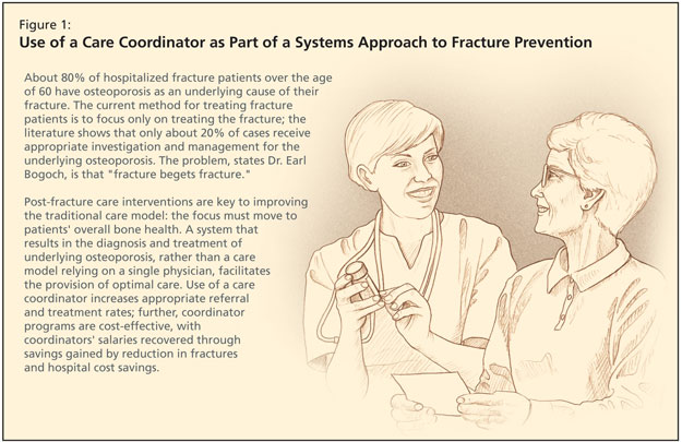


In this program, coordinators screened all orthopedic inpatients as well as outpatients in their fracture clinic, who had low-trauma fractures of the wrist, hip, vertebrae, and humerus. Coordinators assessed the etiology of the osteoporosis and the patient’s fracture risk. They gave patients recommendations, provided educational materials, communicated with their family physicians, and then closely followed every patient.

Three messages were delivered to fragility fracture patients: 1) Your fracture is probably related to underlying weakness of the bone. 2) By having this fracture, you are now at risk of a hip fracture. 3) Treatment is convenient, safe, and effective.

After 1 year, they found that 95% of their patients received appropriate osteoporosis attention. At 6 months and at 1 year, three-quarters of the patients had undergone a BMD as recommended. Three-quarters of those referred to a specialist had attended, and at 1 year, 50% were adherent with medications. Dr. Bogoch noted that more recent data showed closer to 85% adherence.

According to Dr. Bogoch, similar coordinator or fracture liaison service studies have shown a comparable increase in treatment rates. Further, working with a health economist, they found that the coordinator program was cost-effective, with the coordinator’s salary more than recovered. Additionally, in their cohort of 500 patients, predicted hip fractures were reduced, with considerable hospital cost savings.

There were also other benefits to working with a coordinator: orthopedic surgeons were more likely to document information about overall fragility rather than only the fracture; there was an increase in identification of atypical osteoporosis; there was an improvement in patient knowledge and attitudes; and there was an increase in appropriate referrals to osteoporosis specialists.

A commitment for a comprehensive osteoporosis strategy has now been made in Ontario, and $5 million in annual funding has been committed for this project. The program now includes most types of low-trauma fracture, since they now know they are all predictors of a future hip fracture. Nineteen coordinators work in 33 fracture clinics across Ontario.

They began screening patients in early 2007 and results of the first year were reported. The coordinator met with more than 26,000 patients, about 13,000 of whom completed baseline information questionnaires. They educated 12,000 patients and had an intervention with their family doctor; over 10,000 patients were told they should discuss a BMD with their family physician; and 8,700 family doctors received a letter recommending osteoporosis follow-up. These patients will be followed, and their data linked to CIHI and Ontario Health data, for future fracture rates as well as their utilization of osteoporosis medications, in the over-65 group.
Victoria Elliot-Gibson, osteoporosis coordinator at St. Michael’s and program consultant for Osteoporosis Canada, was the original coordinator of the program, and she addressed the symposium. She explained that osteoporosis screening coordinators are hired by Osteoporosis Canada.

All centres follow a protocol to provide uniformity of care. They screen via a variety of methods because coordinators do not have similar access across all sites, using electronic records, paper charts, or ER referrals. The assessment for each patient is done in a private area. Coordinators are also now collecting consent forms so they can use this data for research purposes. Coordinators have a network, with a website, www.OSCnet.ca. It features a forum for questions to Ms. Elliot-Gibson, as well as relevant articles so that coordinators can stay current with the literature.

After the Fall: The ABCs of Fracture Prevention

After the Fall: The ABCs of Fracture Prevention

Teaser: 

Susan B. Jaglal, PhD, Toronto Rehabilitation Institute Chair, Associate Professor, Faculty of Medicine, Department of Physical Therapy, University of Toronto, Toronto, ON.

A wrist fracture is associated with an increased risk of another fracture and should prompt investigation for osteoporosis in both men and women. If the fracture was caused by low trauma (a fall from a standing height or less), a bone density test should be ordered. If the T score is <–1.5, pharmacological treatment with a bisphosphonate and calcium (1,500 mg/d) and vitamin D3 (≥800 IU/d) is recommended. Management should also include balance, posture, and muscle-strengthening exercises and walking, as well as a review of fall-prevention strategies.
Key words: wrist fracture, osteoporosis, diagnosis, treatment, exercise, falls.

Vertebral Compression Fractures Among Older Adults

Vertebral Compression Fractures Among Older Adults

Teaser: 

Simona Abid, MD, FRCP(C), Geriatric Medicine Fellow, McMaster University, Hamilton, ON.
Alexandra Papaioannou, MD, FRCP(C) MSc, Professor, Department of Medicine, Division of Geriatric Medicine, McMaster University, Hamilton, ON.

Vertebral compression fractures (VCF) are the hallmark of osteoporosis, yet two-thirds of all VCF remain undiagnosed and untreated. Both symptomatic and occult VCF are associated with considerable increases in morbidity and mortality, hospitalization rates, admissions to long-term care, and health care-related costs. These fractures increase the risk of future osteoporotic fractures, both vertebral and nonvertebral, independent of bone mineral density. Older adults have lower rates of diagnosis and treatment compared with younger patients, although clinical studies have shown the efficacy and safety of currently available therapies for osteoporosis in older adults are comparable with those in younger individuals.
Key words: vertebral compression fractures, osteoporosis, bone mineral density, antiresorptive therapy, anabolic agents.

New Pharmacotherapy for Osteoporosis

New Pharmacotherapy for Osteoporosis

Teaser: 

Savannah Cardew, MD, FRCP(C), Osteoporosis Program, University Health Network and Mount Sinai Hospital, University of Toronto, Toronto, ON.

Successful management of osteoporosis includes nonpharmacologic and pharmacologic strategies, aimed at fracture prevention. First-line therapies include oral bisphosphonates, an intravenous bisphosphonate (zoledronic acid) that is administered once yearly, the selective estrogen receptor modulator raloxifene and parathyroid hormone. Other selective estrogen receptor modulators are being investigated as potential therapies. Strontium ranelate and denosumab each have a unique mechanism of action and may eventually be available in Canada for the management of osteoporosis. In this article the aforementioned therapies will be reviewed with an emphasis on their efficacy in preventing fractures.
Key words: osteoporosis, osteoporotic fractures, zoledronic acid, parathyroid hormone, raloxifene.

Osteoporosis Screening and Assessment of Fracture Risk

Osteoporosis Screening and Assessment of Fracture Risk

Teaser: 


Mohammed O. Rahman, BHSc student, McMaster University, Hamilton, ON.
Aliya Khan, MD, FRCPC, FACP, FACE, Professor of Clinical Medicine, McMaster University, Hamilton, ON, Director, Calcium Disorders Clinic, St. Joseph’s Healthcare, Hamilton; Director, Oakville Bone Center, Oakville, ON.

Osteoporosis is a skeletal disease characterized by impaired bone strength and an increased risk of fragility fracture. Effective screening should be aimed at evaluating risk factors for osteoporosis with identification of individuals at risk, allowing for intervention prior to fragility fracture. This article presents an overview of the risk factors for fracture in men and women and the integration of these factors in various models, enabling an assessment of the 10-year fracture risk. Through effective screening, early identification, and early intervention with pharmacological therapy of osteoporosis, significant impact can be made on reducing fragility fracture incidence, thereby alleviating the economic and clinical costs to our health care system.
Key words: osteoporosis, screening, risk factors, diagnosis, FRAX.

Osteoporosis Fracture Prevention in Long-Term Care

Osteoporosis Fracture Prevention in Long-Term Care

Teaser: 


Cathy R. Kessenich, DSN, ARNP, Professor of Nursing, University of Tampa, Tampa, FL, USA.
Darlene A. Higgs, RN, BSN, Nurse Practitioner Student, University of Tampa, Tampa, FL, USA.

Osteoporosis is a major cause of health problems in residents of long-term care facilities. It often results in bone fracture, causing poor quality of life and a national financial burden. As the population ages, the incidence of osteoporosis and its consequences increase. It is essential to employ fracture-prevention strategies that have proven most beneficial in long-term care settings and those tailored to promote adherence among older adults. This article reviews osteoporotic treatment appropriate for individuals in long-term care, including treatment through pharmacology, nutritional support, fall prevention, and hip fracture prevention.
Key words: osteoporosis, long-term care, hip protectors, fall prevention, vitamin D.

Continuing the Effort to Improve Outcomes||in Osteoporosis among Aging Adults

Continuing the Effort to Improve Outcomes||in Osteoporosis among Aging Adults

Teaser: 

Since our last issue focusing on osteoporosis, much has changed and yet much stays the same. We have new Canadian guidelines on the detection and management of osteoporosis, several new treatment modalities, and a better understanding of osteoporosis in men. However, many people with osteoporotic fractures are not identified and managed, many people do not stay on effective treatment, and falls prevention programs are rarely available for appropriate older adults. In other words, we have a long way to go before we can say we are dramatically ameliorating the morbidity that osteoporosis causes. Hopefully, this edition of Geriatrics & Aging will do its part to promote the effective diagnosis and management of this important disorder.

Two articles tackle the important issue of diagnosing osteoporosis and determining who is at particular risk for complications. Dr. Angela Juby and Dr. David Hanley review “Diagnostic Tools for Osteoporosis in Older Adults” while Dr. Aliya Khan and Mohammed Rahman discuss one of the key issues in their article “Osteoporosis Screening and Assessment of Fracture Risk.” In our CME article, Dr. Savannah Cardew reviews “New Pharmacotherapy for Osteoporosis.” Our final focus article concentrates on a group at high risk of having osteoporosis and not being treated. Cathy Kessenich and Darlene Higgs review “Osteoporosis Fracture Prevention in Long Term Care.”

We also have our usual collection of key geriatric topics. My colleague, Dr. M. Bachir Tazkarji reviews the important area of “Blood Pressure and Cardiovascular Risk among Older Adults.” Dr. Ekaterina Rogaeva reviews an area of intense research scrutiny in her article on “The Genetic Profile of Alzheimer’s Disease: Updates and Considerations.” Dr. Sophie Robichaud and Dr. Joseph Blondeau address a common problem in their article “Urinary Tract Infections in Older Adults: Current Issues.” Educating students, residents, and even ourselves in proper medical care of the older adult has been a tremendous challenge so any improvements offered via technology are much appreciated. In their article, Drs. Anita Bagri, Bernard Roos, and Jorge Ruiz discuss “Simulation Technology in Geriatric Education.” And our Sexual Health column, written by Drs. Irwin Kuzmarov and Jerald Bain of our partner organization the Canadian Society for the Study of the Aging Male, looks at the important matter of "Sexuality and the Aging Couple, Part I: The Aging Woman."

Enjoy this issue,
Barry Goldlist