Advertisement

Advertisement

fracture

Spinal Injuries among Paediatric Patients

Teaser: 

Dr. Khaled Almansoori, MD, M.Ed, FRCSC,

Adult & Paediatric Spine Surgeon, Department of Orthopaedic Surgery, Advocate Christ Medical Center, Illinois, USA.

CLINICAL TOOLS

Abstract:Due to the distinctive anatomic and biomechanical features of the growing paediatric spine, children are susceptible to unique patterns of spinal injuries. Although clinical examination can help guide management, physicians are often required to rely on advanced imaging. Imaging interpretation can be challenging when considering that abnormal parameters among adults, are often within normal physiological limits in children. In general, spinal injuries in children younger than nine years of age are often managed non-operatively, while adolescents are typically managed by adult treatment principles. With the exception of neurologic injuries, most paediatric spinal injuries demonstrate good to excellent prognosis and outcomes.
Key Words: fracture, injury, spine, paediatric, children.

Members of the College of Family Physicians of Canada may claim MAINPRO-M2 Credits for this unaccredited educational program.

www.cfpc.ca/Mainpro_M2

You can take quizzes without subscribing; however, your results will not be stored. Subscribers will have access to their quiz results for future reference.

Due to the unique properties of the growing spine, including greater elasticity, osseous plasticity, presence of growth centers, relatively strong ligaments, and greater joint mobility, paediatric patients are susceptible to unique fracture patterns and injuries.
There are absolute contraindications regarding return to play decisions.
Children under 13 years of age with vertebral body compression fractures can progressively restore their vertebral height until skeletal maturity.
The vast majority of spine injuries among children under nine years of age, even when relatively unstable, can be managed non-operatively.
Pre-adolescent patients with complete spinal cord injuries are at high risk for developing progressive scoliosis and have not been shown to demonstrate any better neurological outcomes when compared to adults.
The cervical spine is the commonest area of spine injuries with the C1-3 vertebral levels being more commonly seen in children under eight years of age.
A standard immobilization board should not be used for children under eight years of age without an occipital recess or 2-3cm of padding to elevate their body relatively to their head.
Adult radiographic spinal parameters are often unreliable in children and severe neurologic injuries can be sustained in spite of normal imaging results.
Clinical examination is fairly unreliable for identifying spinal column injuries among pre-school patients and it is often necessary to rely on advanced imaging.
To have access to full article that these tools were developed for, please subscribe. The cost to subscribe is $80 USD per year and you will gain full access to all the premium content on www.healthplexus.net, an educational portal, that hosts 1000s of clinical reviews, case studies, educational visual aids and more as well as within the mobile app.
Disclaimer: 
Disclaimer at the end of each page

Approche systémique de la prévention des fractures : le Projet||Ontario

Approche systémique de la prévention des fractures : le Projet||Ontario

Teaser: 

Approche systémique de la prévention des fractures : le Projet Ontario

Conférencier : Earl Bogoch, M.D., FRCSC, Université de Toronto ; Hôpital St-Michael’s, Toronto, ON.

Le message principal du Dr Earl Bogoch a porté sur l’importance de déterminer et de traiter l’ostéoporose après une fracture de fragilité, afin de prévenir de futures fractures, surtout celles de la hanche.

Le manque de soins aux patients après une fracture de fragilité est un fait bien connu qu’on retrouve partout dans le monde. C’est un problème important ; l’ostéoporose est une cause sous-jacente des fractures chez environ 80 % des patients de plus de 60 ans hospitalisés pour fracture. À partir de 50 ans, la moitié des femmes et un cinquième des hommes risquent d’avoir une fracture ostéoporotique. Le fardeau économique a été bien établi, tout comme la morbidité—et la mortalité—individuelle importante, chez ceux qui ont des fractures de la hanche, avec perte de fonction et d’indépendance.

La méthode actuelle pour traiter les patients ayant une fracture de fragilité peut ne consister qu’en un traitement de la fracture ; la littérature médicale indique que seuls environ 20 % des cas reçoivent l’attention et la prise en charge appropriées à l’ostéoporose sous-jacente.

Le Dr Bogoch a fait la critique du modèle traditionnel de soins qui ne repose que sur l’expertise et l’initiative d’un seul médecin, ce qui joue un piètre rôle dans les soins de l’ostéoporose. Les chirurgiens s’occupent des fractures, mais souvent personne ne fait attention à la santé osseuse des patients. La solution qu’il a proposée consiste en un système conduisant au diagnostic et au traitement, plutôt qu’en un modèle de soins ne reposant que sur un seul médecin.

Il y a dix ans, le Dr Bogoch a mené une étude dans cinq hôpitaux ontariens. Les patients ayant eu des fractures du poignet, de l’épaule, des vertèbres ou de la hanche ont reçu une explication par écrit sur les risques de l’ostéoporose, et une lettre a été envoyée au médecin de famille recommandant un suivi de l’ostéoporose. Par la suite, moins des deux tiers ont consulté leur médecin de famille et, parmi ceux-ci, seuls 69 % ont eu une densitométrie. Le taux de traitement n’a que très peu augmenté, de 17 % (groupe témoin historique) à 24 %. Déçu par les résultats, le Dr Bogoch en a conclu qu’il ne suffisait pas de simplement informer les patients et les médecins ; il leur fallait un coordinateur pour promouvoir les soins appropriés.

D’autres études en Ontario ont donné des résultats similaires ; les taux de densitométrie et de traitement n’ont augmenté que de façon marginale, voire pas du tout. Le manque de succès de ces programmes a conduit à la décision de mettre l’accent sur le programme de coordinateur (Figure 1).


Dans ce programme, les coordinateurs ont évalué tous les patients orthopédiques hospitalisés ainsi que les patients externes dans leur clinique de fractures, patients qui avaient subi des fractures peu traumatiques du poignet, de la hanche, des vertèbres ou de l’humérus. Les coordinateurs ont évalué l’étiologie de l’ostéoporose et le risque de fracture des patients. Ils ont fait des recommandations aux patients, leur ont fourni du matériel didactique, ont communiqué avec leur médecin de famille et ont aussi suivi chaque patient de près.
Trois messages ont été adressés aux patients ayant eu une fracture de fragilité : 1) Votre fracture est probablement liée à une fragilité sous-jacente de l’os. 2) Ayant eu cette fracture, vous risquez maintenant une fracture de la hanche. 3) Le traitement est facile, sécuritaire et efficace.

Après un an, ils ont constaté que 95 % de leurs patients avaient reçu l’attention appropriée à leur ostéoporose. À 6 mois et à 1 an, trois quarts des patients avaient reçu le test de la DMO recommandé. Trois quarts de ceux qui avaient été adressés à un spécialiste l’avaient consulté et, au bout d’un an, 50 % respectaient leur médication. Le Dr Bogoch a signalé que des données plus récentes faisaient état de près de 85 % d’observance thérapeutique.

Selon le Dr Bogoch, des études similaires avec coordinateurs ou services de liaison pour les fractures ont indiqué une hausse comparable des taux de traitement. De plus, en travaillant avec un économiste de la santé, ils ont démontré que le programme de coordination était rentable, le salaire du coordinateur étant plus que compensé. En outre, dans leur cohorte de 500 patients, les fractures de la hanche prédites avaient diminué, induisant des économies considérables de frais d’hôpitaux.

Il y a aussi eu d’autres avantages à travailler avec un coordinateur : les chirurgiens orthopédiques étaient plus enclins à documenter les données sur la fragilité générale plutôt que de se limiter à la fracture ; il en a résulté une augmentation d’identification de l’ostéoporose atypique ; il y a eu une amélioration de la connaissance et de l’attitude des patients ; et il y a eu une augmentation des orientations appropriées vers les spécialistes de l’ostéoporose.

L’Ontario a maintenant pris un engagement pour une stratégie étendue de l’ostéoporose, et un budget annuel de 5 millions de dollars a été alloué à ce projet. Le programme comprend maintenant la plupart des types de fractures peu traumatiques, car on sait dorénavant qu’elles sont toutes des prédicteurs d’une future fracture de la hanche. Dix-neuf coordinateurs travaillent dans 33 cliniques des fractures à travers l’Ontario.
Ils ont commencé à évaluer les patients au début de 2007 et les résultats de la première année sont disponibles. Les coordinateurs ont vu plus de 26 000 patients, dont près de 13 000 ont répondu à des questionnaires de renseignements de référence. Ils ont éduqué 12 000 patients et sont intervenus auprès de leur médecin de famille ; ils ont conseillé plus de 10 000 patients d’aborder la DMO avec leur médecin de famille ; et 8 700 médecins de famille ont reçu une lettre recommandant un suivi de l’ostéoporose. Ces patients seront suivis, et leurs données seront associées aux données de l’ICIS et de Santé Ontario pour le calcul de futurs taux de fracture et pour connaître leur utilisation des médicaments contre l’ostéoporose, dans le groupe des plus de 65 ans.

Victoria Elliot-Gibson, coordinatrice pour l’ostéoporose à l’hôpital St-Michael’s et consultante du programme Ostéoporose Canada, a été la coordinatrice initiale du programme, et elle s’est adressée aux participants du symposium. Elle a expliqué que les coordinateurs du dépistage de l’ostéoporose sont engagés par Ostéoporose Canada.

Tous les centres suivent un protocole pour dispenser des soins uniformes. Ils réalisent les dépistages grâce à diverses méthodes, car les coordinateurs n’ont pas les mêmes moyens dans tous les sites, se servant de dossiers électroniques, de graphes sur papier ou d’orientation aux urgences. L’évaluation de chaque patient est effectuée en zone privée. Les coordinateurs recueillent maintenant aussi des formulaires de consentement pour pouvoir utiliser les données à des fins de recherche. Les coordinateurs ont un réseau, dont le site Web est le www.OSCnet.ca. On y trouve un forum pour les questions à Mme Elliot-Gibson, ainsi que des articles pertinents pour que les coordinateurs puissent demeurer à jour dans leur domaine.

Systems Approach to Fracture Prevention: The Ontario Project

Systems Approach to Fracture Prevention: The Ontario Project

Teaser: 


Systems Approach to Fracture Prevention: The Ontario Project

Speaker: Earl Bogoch, MD, FRCSC, University of Toronto; St. Michael’s Hospital, Toronto, ON.

Dr. Earl Bogoch’s key message concerned the importance of identifying and treating osteoporosis after a fragility fracture, in order to prevent future fractures, most importantly of the hip.

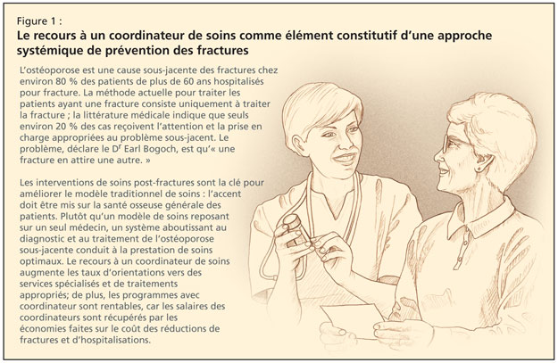
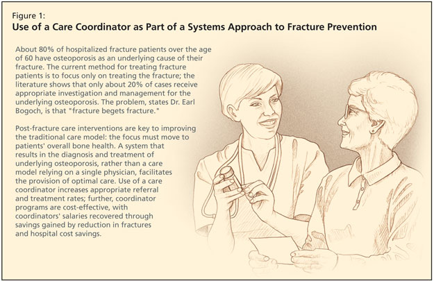
The care gap for patients after a fragility fracture is well-known and exists internationally. This problem is significant; about 80% of hospitalized fracture patients over the age of 60 have osteoporosis as an underlying cause of their fracture. From the age of 50, half of women and one-fifth of men are likely to experience an osteoporotic fracture. The economic burden has been well documented as well as major personal morbidity—and mortality—in those with hip fractures, with loss of function and independence.

The current method for treating fragility fracture patients may focus only on treating the fracture; the literature shows that only about 20% of cases receive appropriate investigation and management for the underlying osteoporosis.

Dr. Bogoch offered a critique of the traditional care model that relies on the expertise and initiative of a single doctor, which serves osteoporosis care poorly. While surgeons deal with fractures, frequently no one focuses on patients’ bone health. The solution he posed is a system that results in the diagnosis and treatment, rather than a care model relying on a single physician.

Ten years ago, Dr. Bogoch carried out a study in five Ontario hospitals. Patients with wrist, shoulder, vertebral, or hip fractures were provided with a written explanation of the risks of osteoporosis, and a letter was mailed to the family doctor recommending osteoporosis follow-up. Afterward, less than two-thirds saw their family doctor and, of those, 69% had densitometry. Treatment rate went up only slightly, from 17% (historical controls) to 24%. Disappointed with the results, Dr. Bogoch determined that simply informing patients and physicians was insufficient; they needed a coordinator to promote good care.

Other studies in Ontario had similar results; densitometry and treatment rates increased minimally or not at all. The lack of success with these programs led to their decision to focus on the coordinator program (Figure 1).


In this program, coordinators screened all orthopedic inpatients as well as outpatients in their fracture clinic, who had low-trauma fractures of the wrist, hip, vertebrae, and humerus. Coordinators assessed the etiology of the osteoporosis and the patient’s fracture risk. They gave patients recommendations, provided educational materials, communicated with their family physicians, and then closely followed every patient.

Three messages were delivered to fragility fracture patients: 1) Your fracture is probably related to underlying weakness of the bone. 2) By having this fracture, you are now at risk of a hip fracture. 3) Treatment is convenient, safe, and effective.

After 1 year, they found that 95% of their patients received appropriate osteoporosis attention. At 6 months and at 1 year, three-quarters of the patients had undergone a BMD as recommended. Three-quarters of those referred to a specialist had attended, and at 1 year, 50% were adherent with medications. Dr. Bogoch noted that more recent data showed closer to 85% adherence.

According to Dr. Bogoch, similar coordinator or fracture liaison service studies have shown a comparable increase in treatment rates. Further, working with a health economist, they found that the coordinator program was cost-effective, with the coordinator’s salary more than recovered. Additionally, in their cohort of 500 patients, predicted hip fractures were reduced, with considerable hospital cost savings.

There were also other benefits to working with a coordinator: orthopedic surgeons were more likely to document information about overall fragility rather than only the fracture; there was an increase in identification of atypical osteoporosis; there was an improvement in patient knowledge and attitudes; and there was an increase in appropriate referrals to osteoporosis specialists.

A commitment for a comprehensive osteoporosis strategy has now been made in Ontario, and $5 million in annual funding has been committed for this project. The program now includes most types of low-trauma fracture, since they now know they are all predictors of a future hip fracture. Nineteen coordinators work in 33 fracture clinics across Ontario.

They began screening patients in early 2007 and results of the first year were reported. The coordinator met with more than 26,000 patients, about 13,000 of whom completed baseline information questionnaires. They educated 12,000 patients and had an intervention with their family doctor; over 10,000 patients were told they should discuss a BMD with their family physician; and 8,700 family doctors received a letter recommending osteoporosis follow-up. These patients will be followed, and their data linked to CIHI and Ontario Health data, for future fracture rates as well as their utilization of osteoporosis medications, in the over-65 group.
Victoria Elliot-Gibson, osteoporosis coordinator at St. Michael’s and program consultant for Osteoporosis Canada, was the original coordinator of the program, and she addressed the symposium. She explained that osteoporosis screening coordinators are hired by Osteoporosis Canada.

All centres follow a protocol to provide uniformity of care. They screen via a variety of methods because coordinators do not have similar access across all sites, using electronic records, paper charts, or ER referrals. The assessment for each patient is done in a private area. Coordinators are also now collecting consent forms so they can use this data for research purposes. Coordinators have a network, with a website, www.OSCnet.ca. It features a forum for questions to Ms. Elliot-Gibson, as well as relevant articles so that coordinators can stay current with the literature.

Osteoporosis Screening and Diagnosis

Osteoporosis Screening and Diagnosis

Teaser: 


Rowena Ridout, MD, FRCPC, Toronto Western Hospital, Toronto, ON.

Osteoporosis is a significant cause of morbidity and mortality in the older population. Bone density testing is recommended for all men and women 65 or older. In postmenopausal women, and in men over the age of 50, testing is recommended for those at high risk for osteoporosis. Effective therapy is available for those at risk for fracture. Bone density testing combined with clinical risk factors, including age and fracture history, can be used to assess fracture risk and identify those individuals most likely to benefit from drug therapy.
Key words: osteoporosis, bone density, fracture, diagnosis.

Osteoporosis in Men: Myth or Fact

Osteoporosis in Men: Myth or Fact

Teaser: 

Wojciech P.Olszynski, MD, PhD, FRCPC, Clinical Professor of Medicine,University of Saskatchewan, Director, Saskatoon Osteoporosis Centre, Saskatoon, SK.

Though osteoporosis occurs less frequently in men than in women, it is nonetheless a significant medical problem. Osteoporotic vertebral fractures in particular are as common for men as for women, and about one-third of all hip fractures occur in men. As a consequence of fragility fractures, the associated morbidity and mortality are higher in men than women, particularly after fracture of the hip. Idiopathic osteoporosis is common; however, secondary causes are found in about 50% of cases. Bone density measurements should be advised for every man over 65 years of age and for younger men in the presence of osteoporosis risk factors. For practical purposes, the use of T-score <= 2.5 for men over age 65 should be used for the diagnosis of osteoporosis.

Key words: osteoporosis, men, fracture, diagnosis, treatment

Epidemiology
Osteoporosis is a skeletal disorder characterized by compromised bone strength predisposing a person to an increased risk of fracture.

Osteopenia--Fracture Risk Doubles Every Decade Past Age 50

Osteopenia--Fracture Risk Doubles Every Decade Past Age 50

Teaser: 

Shechar Dworski, BSc

Osteopenia literally means "poverty of bone," while osteoporosis (OP) means "porous bone." The underlying cause of both conditions is a difference in the rate of bone formation and bone loss. Normally, both processes take place at equal rates resulting in a dynamic equilibrium. Bone density peaks during the second or third decade of life and then gradually declines with age, when bone loss exceeds bone formation. Bone is formed in response to physical stresses imposed on it, so excessive loss may occur as a result of immobility. Other causes of excessive loss include hormonal changes, either after menopause, or with excess parathyroid or corticosteroid hormones, or insufficient vitamin D or calcium intake.

In radiological terms, osteopenia refers to an increased radiolucency of bone. The most common cause of this is OP, although there are other causes for osteopenia, such as osteomalacia (so-called "renal rickets", Vitamin D deficiency-related problems), hyperparathyroidism, and some renal diseases. Renal osteodystrophy (or uremic bone disease) is the term for a complex group of bone disorders that occur in patients with chronic renal failure (CRF). Specific radiographic clues for other causes of osteopenia include: looser zones found in osteomalacia, subperiosteal resorption present in hyperparathyroidism, and focal lytic lesions (as seen in disseminated multiple myeloma).