Advertisement

Advertisement

telomere shortening

Télomères, télomérase et vieillissement

Télomères, télomérase et vieillissement

Teaser: 

Télomères, télomérase et vieillissement

Conférencière : Chantal Autexier, Ph. D., Institut Lady Davis de recherches médicales, Hôpital général juif et Centre Bloomfield de recherche sur le vieillissement; départements de médecine et d’anatomie et de biologie cellulaire, Université McGill, Montréal (Québec).

La Dre Chantal Autexier a discuté du rôle des télomères dans le maintien de l’intégrité génétique et cellulaire, et du rôle de la perturbation des télomères dans la sénescence cellulaire et le vieillissement.

Télomères : description et rôle dans l’intégrité cellulaire
Les télomères sont des séquences répétitives d’ADN non codant (TTAGGG) qui protègent les extrémités des chromosomes eucaryotes linéaires. Chez les hommes, un complexe shelterin, formé de six protéines, se lie aux télomères1. La structure et la taille des télomères sont importantes pour le maintien de l’intégrité cellulaire (Figure 1). Les télomères empêcheraient les mécanismes de réponse aux lésions cellulaires de considérer les extrémités des chromosomes linéaires comme des cassures d’ADN double brin (notamment comme des lésions chromosomiques provoquées par des stress, tels que des radiations ionisantes). Une perturbation de la structure ou de la taille du télomère peut entraîner des fusions entre les extrémités des chromosomes, une instabilité et des anomalies chromosomiques, et des troubles de la division cellulaire (sénescence cellulaire; mort cellulaire; les cellules deviennent cancéreuses).

La majorité des cellules de l’orga-nisme sont incapables de conserver la taille du télomère d’une division à l’autre, car la machine de réplication de l’ADN est incapable de répliquer la totalité des extrémités chromosomiques. Par conséquent, le télomère se raccourcit avec chaque division cellulaire. Une fois une certaine limite atteinte (la limite de Hayflick), la majorité des cellules sortent du cycle cellulaire et rentrent en sénescence. Cependant, une petite proportion de cellules est capable de réintégrer le cycle cellulaire : ce phénomène est en général associé à une élongation des télomères et à une expression de la télomérase, une enzyme impliquée dans la formation et le maintien des télomères. Bodnar et ses collègues ont montré qu’une expression forcée de la télomérase inhibe la perte des télomères et l’arrêt de la croissance de fibroblastes humains normaux en culture, et suffit à l’élongation du télomère et à l’immortalisation cellulaire (prolongation de la durée de vie)2. À la différence de la majorité des cellules, certaines cellules, comme les cellules souches, les cellules de la lignée germinale et 85 % des cellules cancéreuses, expriment la télomérase. Par conséquent, ces cellules sont capables de maintenir la taille et la structure des télomères durant un grand nombre de cycles de division cellulaire.

Raccourcissement des télomères, sénescence cellulaire et vieillissement tissulaire
On appelle sénescence réplicative le processus par lequel une cellule sort du cycle cellulaire. Ce processus peut être déclenché par différents stress, comme des lésions dues aux radiations ou un stress oxydatif3,4. Dans de nombreux cas, une perte de l’intégrité des télomères est responsable de la sénescence ou y est associée.

Le cycle cellulaire est contrôlé par de multiples mécanismes de vérification qui détectent les lésions de l’ADN, activent les mécanismes de réparation et déclenchent la mort cellulaire lorsque les lésions ne peuvent être réparées. Les suppresseurs de tumeurs sont d’importants régulateurs du cycle cellulaire. Ils régulent la sénescence et la mort cellulaire, veillant à ce que toute cellule très abîmée meurt ou arrête de se diviser. Lorsque ces protéines de contrôle sont absentes ou ne fonctionnent pas correctement, les cellules porteuses de lésions continuent à se développer. Bien que les suppresseurs de tumeurs protègent un organisme, en empêchant l’accumulation de cellules abîmées, ils contribuent également au vieillissement tissulaire. En effet, à mesure que les cellules abîmées qui rentrent en sénescence sont éliminées, le pool des cellules responsables du renouvellement des tissus et du maintien de la fonction tissulaire diminue.

Chez l’homme et d’autres organismes, les tissus qui se renouvellent rapidement (paroi de l’intestin, peau, follicules pileux et moelle osseuse) sont les premiers affectés par le raccourcissement des télomères et la sénescence et la mort cellulaires. De nombreux troubles liés au vieillissement affectent ces tissus.

Selon la Dre Autexier, de nombreuses études ont montré une corrélation entre le raccourcissement des télomères et la sénescence réplicative, la mort cellulaire et le vieillissement, d’une part, et entre la préservation de la taille des télomères, la présence d’une activité télomérase et l’immortalisation cellulaire, la longévité ou la formation d’un cancer, d’autre part.

Perturbation des télomères et vieillissement
La Dre Autexier a expliqué que de nombreuses caractéristiques du vieillissement naturel chez l’homme sont liées à un déclin des mécanismes nécessitant un renouvellement cellulaire constant, ou à un défaut des mécanismes de protection empêchant l’apparition ou la survie de cellules présentant des anomalies génomiques. Il s’agit notamment d’une diminution des fonctions immunitaires et de la moelle osseuse, d’une modification de l’épaisseur de la peau, d’une moins bonne cicatrisation des blessures, de modifications structurales des tissus épithéliaux, d’une perte de la fécondité et d’une augmentation de l’incidence du cancer. Les recherches suggèrent maintenant qu’une perte de l’intégrité des télomères est l’un des mécanismes clés à l’origine des pathologies liées à l’âge et au vieillissement.

La Dre Autexier a expliqué qu’il existe des similarités phénotypiques entre des syndromes de vieillissement prématuré chez l’homme, comme l’ataxie- télangiectasie, le syndrome de Werner et la dyskératose congénitale (DC), et le vieillissement normal, notamment des cataractes, une ostéoporose, un grisonnement des cheveux et une neurodégénérescence5. Ces syndromes sont associés à une défectuosité des gènes jouant un rôle dans le maintien de l’intégrité génomique et télomérique. Les phénotypes associés à la DC s’observent d’abord dans les tissus soumis à un renouvellement cellulaire constant, comme l’intestin, l’épiderme et la moelle osseuse6,7. Les populations de patients atteints d’une forme dominante autosomique de la maladie ont des télomères plus courts, montrent des signes de la maladie plus tôt et une aggravation des symptômes à chaque génération successive. Les résultats sont en accord avec un mécanisme défectueux de maintien des télomères.

Effectivement, diverses formes de DC dérivent de mutations touchant des gènes directement impliqués dans le maintien de l’intégrité des télomères. À ce jour, les mutations identifiées comme étant associées à la DC affectent la télomérase, un élément du complexe télomérase appelé hTR (human Telo-merase RNA; ARN de la télomérase humaine) et un gène codant pour la dyskérine, une protéine qui se lie à l’hTR8,9.

Un modèle murin dans lequel le gène mTR a été inactivé a permis de confirmer la relation entre une activité défectueuse de la télomérase et le processus de vieillissement. Bien que ces souris fussent viables, les générations successives montraient un raccourcissement progressif des télomères et une diminution de la fertilité (la 6e génération étant infertile)10. Il est intéressant de noter que ces souris présentent également une mauvaise cicatrisation, une perte et un grisonnement des poils, une diminution du poids corporel, une atrophie des villosités intestinales, une insuffisance médullaire et une augmentation de l’incidence du cancer11,12.

La majorité des phénotypes observés dans ce modèle murin sont semblables à ceux des patients atteints de DC et, à un certain point, ressemblent aux pathologies liées à l’âge associées au vieillissement humain normal13.

Conclusion
La Dre Autexier a terminé sa présentation en observant que de nombreuses études au cours de ces quatre dernières années corrélaient la taille des télomères à la mortalité, la maladie d’Alzheimer et la longévité. Bien que ces études soient généralement des études de corrélation, il est intéressant de voir que la recherche s’intéresse à la longueur des télomères comme marqueur potentiel de certaines maladies associées au vieillissement14,15,16.

L’intégrité des télomères est un régulateur essentiel du vieillissement humain et de la formation de cancers. Cette observation suggère que des protéines impliquées dans le maintien des télomères pourraient représenter des cibles moléculaires d’interventions thérapeutiques dans le domaine du cancer et d’autres maladies liées au vieillissement.

Bibliographie

  1. Blasco MA. Telomere length, stem cells and aging. Nat Chem Biol 2007;3:640-9.
  2. Bodnar AG, Ouellette M, Frolleis M, et al., Extension of life-span by introduction of telomerase into normal human cells. Science 1998;279:349-52.
  3. Campisi J. Senescent cells, tumor suppression, and organismal aging: good citizens, bad neighbors. Cell 2005;120:513-22.
  4. Ithana K, Campisi J, Dimri GP. Mechanisms of cellular senescence in human and mouse cells. Biogerontol 2004;5:1-10.
  5. Hasty P, Campisi J, Hoeijmakers J, et al. 2003. Aging and genome maintenance: lessons from the mouse? Science 2003;299:1355-9.
  6. Kirwan M, Dokal I. Dyskeratosis congenita: a genetic disorder of many faces. Clin Genet 2008;73:103-12.
  7. Marciniak R, Guarente L. Human genetics. Testing telomerase. Nature 2001;413:370-3.
  8. Mitchell JR, Wood E, Collins K. A telomerase component is defective in the human disease dyskeratosis congenital. Nature 1999;402:551-5.
  9. Dokal I, Vulliamy T. Dyskeratosis congenita: its link to telomerase and aplastic anaemia. Blood Rev 2003;17:217-25.
  10. Blasco MA, Lee HW, Hande MP, et al. Telomere shortening and tumor formation by mouse cells lacking telomerase RNA. Cell 1997;91:25-34.
  11. Rudolph KL, Chang S, Lee HW, et al. Longevity, stress response, and cancer in aging telomerase-deficient mice. Cell 1999;96:701-12.
  12. Lee HW, Blasco MA, Gottlieb GJ, et al. Essential role of mouse telomerase in highly proliferative organs. Nature 1998;392:569-74.
  13. Marciniak RA, Johnson FB, Guarente L. Dyskeratosis congenita, telomeres and human ageing. Trends Genet 2000;16:193-5.
  14. Cawthon RM, Smith, KR, O’Brian E, et al. Association between telomere length in blood and mortality in people aged 60 years or older. Lancet 2003;361:393-5.
  15. Panossian LA, Porter VR, Valenzuela HF, et al. Telomere shortening in T cells correlates with Alzheimer’s disease status. Neurobiol Aging 2003;24:77-84.
  16. Nakamura K, Takubo K, Izumiyama-Shimomura N, et al. Telomeric DNA length in cerebral gray and white matter is associated with longevity in individuals aged 70 years or older. Experimental Gerontology 2007;42:944-50.

Telomeres, Telomerase and Aging

Telomeres, Telomerase and Aging

Teaser: 


Click here to view the entire report from the 28th Annual Scientific Meeting of the Canadian Geriatrics Society

Telomeres, Telomerase and Aging

Speaker: Chantal Autexier, Ph.D., Lady Davis Institute for Medical Research, Jewish General Hospital, and Bloomfield Center for Research in Aging; Departments of Anatomy and Cell Biology, and Medicine, McGill University, Montreal QC.

Dr. Chantal Autexier discussed the role of telomeres in the maintenance of genetic and cellular integrity, and how telomere disruption is involved in cellular senescence and the aging process.

Telomeres and Their Role in Cellular Integrity
Telomeres are noncoding repetitive DNA sequences (TTAGGG) that protect the ends of linear eukaryotic chromosomes. In humans, a shelterin complex of 6 main proteins binds telomeres.1 Both the structure and the length of telomeres are important for cellular integrity (Figure 1).
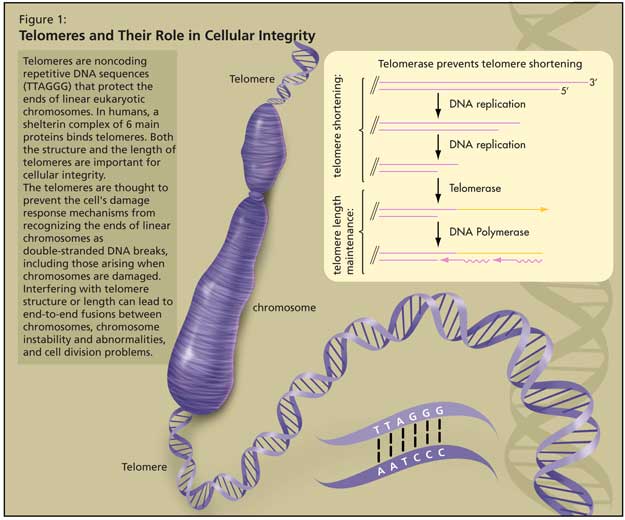
The telomeres are thought to prevent the cell’s damage response mechanisms from recognizing the ends of linear chromosomes as double-stranded DNA breaks, including those arising when chromosomes are damaged by stresses such as ionizing radiation. Interfering with telomere structure or length can lead to end-to-end fusions between chromosomes, chromosome instability and abnormalities, and cell division problems (cell senescence, cell death, or cells becoming cancerous).

In the body, most cells are unable to maintain telomere length from one division to the next, as the DNA replication machinery is unable to fully replicate the ends of chromosomes, leading to telomere shortening every time cells divide. Once a critical point—the Hayflick limit—is reached, most cells exit the cell cycle and undergo cell senescence. However, a small proportion of cells are able to re-enter the cell cycle, and this is usually associated with the lengthening of telomeres and the expression of telomerase, an enzyme directly involved in telomere formation and maintenance. Bodnar et al. have shown that forced expression of telomerase prevents telomere loss and growth arrest of normal human fibroblasts in culture and is sufficient for telomere lengthening and cell immortalization (extended lifespan).2

While most cells do not express telomerase, some cells do, such as stem cells, cells of the germ line, and 85% of cancer cells. Such cells are therefore able to maintain telomere length and structure through a large number of cell division cycles.

Telomere Shortening, Cell Senescence, and Tissue Aging
The process by which a cell exits the cell cycle is called replicative senescence. This can be triggered by various stresses, such as radiation damage and oxidative stress.3,4 In many cases, senescence is caused by, or associated with, loss of telomere integrity.

The cell cycle is controlled by multiple checkpoint mechanisms that sense DNA damage, activate repair mechanisms, and trigger cell death when damage cannot be repaired. Tumour suppressors are important cell cycle regulators that regulate cell senescence and death, ensuring that a cell with extensive damage dies or stops dividing. When these checkpoint proteins are absent or malfunctioning, damaged cells continue to grow. Although tumour suppressors protect an organism from the accumulation of damaged cells, they also contribute to tissue aging. As damaged and senescent cells are eliminated through cell death, cell pools responsible for tissue renewal and the maintenance of tissue function are depleted.

In humans and other organisms, tissues that undergo rapid self-renewal—lining of the gut, skin, hair follicles, and bone marrow—are the ones most affected by telomere shortening, cell senescence and cell death. Many age-related pathologies affect these tissues.

According to Dr. Autexier, many studies have shown a correlation between telomere shortening and replicative senescence, cell death and aging, and between telomere length maintenance, the presence of telomerase activity and cellular immortalization, longevity or cancer formation.

Telomere Disruption in Aging
Dr. Autexier explained that many characteristics of natural aging in humans are related to a decline in processes that require continuous cell renewal, or to defective safeguard mechanisms that prevent the occurrence and survival of cells with genomic abnormalities. These include declines in immune and bone marrow functions, skin thickness changes, reduced wound healing capacity, structural changes in epithelial tissues, loss of fertility, and increased incidence of cancer. Research now suggests that loss of telomere integrity is one of the key mechanisms that drive the aging and age-related pathologies.

Dr. Autexier described that premature human aging syndromes, such as Ataxia Telangiectasia, Werner Syndrome, and Dyskeratosis congenita (DKC), share phenotypic similarities with the normal aging process, such as cataracts, osteoporosis, hair graying, and neurodegeneration.5 These syndromes are associated with defects in genes implicated in the maintenance of genomic and telomeric integrity.

DKC phenotypes first present in tissues that undergo constant cell renewal, such as the gut, the epidermis, and the bone marrow.6,7 Patient populations with the autosomal dominant form of the disease demonstrate shorter telomeres, earlier disease onset and more severe symptoms with each successive generation. These results are consistent with a defective telomere maintenance process.

Various forms of DKC indeed arise from mutations in genes that are directly involved in maintaining telomere integrity. So far, DKC mutations have been identified in telomerase, in a component of the telomerase complex called hTR (human Telomerase RNA) and in the gene encoding Dyskerin, a protein that binds to hTR.8,9

The relationship between defective telomerase activity and the aging process was confirmed in the mTR knockout mouse model. Although these mice are viable, successive generations exhibit progressive shortening of telomeres and decreased fertility (the 6th generation being infertile).10 Interestingly, these mice also present reduced wound healing, hair loss and graying, reduced body weight, intestinal villi atrophy, bone marrow failure, and an increased incidence of cancer.11,12

Most phenotypes observed in this mouse model are similar to those seen in DKC patients and, to a certain extent, to age-related pathologies associated with normal human aging.13

Conclusion
Dr. Autexier closed by observing that there have been numerous studies in the last 4 years correlating telomere length and mortality, Alzheimer’s disease status, and longevity. Although these studies are generally correlative, it is interesting to see that more research is being done to look at telomere length as a marker for some diseases associated with aging.14,15,16

Telomere integrity is a key regulator of human aging and cancer formation, suggesting that proteins involved in telomere maintenance could represent molecular targets for therapeutic interventions in the fields of cancer and other age-related diseases.

References

  1. Blasco MA. Telomere length, stem cells and aging. Nat Chem Biol 2007;3:640-9.
  2. Bodnar AG, Ouellette M, Frolkis M, et al., Extension of life-span by introduction of telomerase into normal human cells. Science 1998;279:349-52.
  3. Campisi J. Senescent cells, tumor suppression, and organismal aging: good citizens, bad neighbors. Cell 2005;120:513-22.
  4. Ithana K, Campisi J, Dimri GP. Mechanisms of cellular senescence in human and mouse cells. Biogerontol 2004;5:1-10.
  5. Hasty P, Campisi J, Hoeijmakers J, et al. Aging and genome maintenance: lessons from the mouse? Science 2003;299:1355-9.
  6. Kirwan M, Dokal I. Dyskeratosis congenita: a genetic disorder of many faces. Clin Genet 2008;73:103-12.
  7. Marciniak R, Guarente L. Human genetics. Testing telomerase. Nature 2001;413:370-3.
  8. Mitchell JR, Wood E, Collins K. A telomerase component is defective in the human disease dyskeratosis congenital. Nature 1999;402:551-5.
  9. Dokal I, Vulliamy T. Dyskeratosis congenita: its link to telomerase and aplastic anaemia. Blood Rev 2003;17:217-25.
  10. Blasco MA, Lee HW, Hande MP, et al. Telomere shortening and tumor formation by mouse cells lacking telomerase RNA. Cell 1997;91:25-34.
  11. Rudolph KL, Chang S, Lee HW, et al. Longevity, stress response, and cancer in aging telomerase-deficient mice. Cell 1999;96:701-12.
  12. Lee HW, Blasco MA, Gottlieb GJ, et al. Essential role of mouse telomerase in highly proliferative organs. Nature 1998;392:569-74.
  13. Marciniak RA, Johnson FB, Guarente L. Dyskeratosis congenita, telomeres and human ageing. Trends Genet 2000;16:193-5.
  14. Cawthon RM, Smith, KR, O’Brian E, et al. Association between telomere length in blood and mortality in people aged 60 years or older. Lancet 2003;361:393-5.
  15. Panossian LA, Porter VR, Valenzuela HF, et al. Telomere shortening in T cells correlates with Alzheimer’s disease status. Neurobiol Aging 2003;24:77-84.
  16. Nakamura K, Takubo K, Izumiyama-Shimomura N, et al. Telomeric DNA length in cerebral gray and white matter is associated with longevity in individuals aged 70 years or older. Experimental Gerontology 2007;42:944-50.

Why Do We Age? What Do Dolly’s Telomeres Tell Us?

Why Do We Age? What Do Dolly’s Telomeres Tell Us?

Teaser: 


Two Theories Linking DNA Damage and Aging: Free Radical/Oxidation vs. Telomere Shortening

Ruwaida Dhala-Vakil, BSc, MSc

There are many factors involved in human aging, and significant progress has been made in this field over the last few decades. Recent evidence from cloned calves suggests that scientists may not merely be able to reverse the cellular damage accumulating with age--they may, in fact, be able to prolong cell life. The cells from these cloned animals lived longer in culture and had longer telomeres than their normal counterparts. If this extension of the cellular life span can be translated into longer life for the entire organism, the calves may live fifty percent longer than normal.1 Additionally, studies on antioxidants show that transgenic Drosophila (the fruit fly), which overexpress antioxidant genes, live 34% longer than controls.2 This article will focus on both the free radical/oxidation theory of aging, and the role of telome-rase in aging.

Free Radical/Oxidation Theory of Aging
In 1956, Denham Harman suggested that there is an age-related accumulation of reactive oxygen species (ROS) which causes damage to cellular components. The damage is targeted to the proteins and DNA in the nucleus and mitochondria, as well as to the proteins and lipids in the cell membrane, and the proteins of the cytoplasm. Mitochondrial DNA is located near the inner mitochondrial membrane, close to the sites where free radicals form.