Advertisement

Advertisement

Case Studies

A Facial Rash Recalcitrant to Treatment with Topical Corticosteroids

A Facial Rash Recalcitrant to Treatment with Topical Corticosteroids

Members of the College of Family Physicians of Canada may claim one non-certified credit per hour for this non-certified educational program.

Mainpro+® Overview
Teaser: 

Francesca Cheung, MD CCFP, is a family physician with a special interest in dermatology. She received the Diploma in Practical Dermatology from the Department of Dermatology at Cardiff University in Wales, UK. She is practising at the Lynde Centre for Dermatology in Markham, Ontario and works closely with Dr. Charles Lynde, MD FRCPC, an experienced dermatologist. In addition to providing direct patient care, she acts as a sub-investigator in multiple clinical studies involving psoriasis, onychomycosis, and acne.

Abstract
Periorificial dermatitis is a common eczematous eruption on the face. Clusters of follicular papules, vesicles, and pustules on an erythematous base are usually found in a perioral distribution. Other common locations include the nasolabial folds and periocular area. An underlying cause may not be found in all cases, but the use of topical corticosteroids on the face may precede onset of symptoms. Periorificial dermatitis is diagnosed clinically and no specific investigation is required. Topical anti-inflammatory therapies (such as metronidazole and erythromycin) are appropriate in mild cases. In severe cases, systemic treatments such as tetracycline or one of its derivatives are beneficial. Patients should be warned that symptoms might worsen before improvement is apparent. This complication is more commonly seen when topical corticosteroids are withdrawn.
Keywords: periorificial dermatitis, perioral dermatitis, facial rash, steroid-induced.

Ear Lesions That Recur Every Spring

Ear Lesions That Recur Every Spring

Members of the College of Family Physicians of Canada may claim one non-certified credit per hour for this non-certified educational program.

Mainpro+® Overview
Teaser: 

Francesca Cheung, MD CCFP, is a family physician with a special interest in dermatology. She received the Diploma in Practical Dermatology from the Department of Dermatology at Cardiff University in Wales, UK. She is practising at the Lynde Centre for Dermatology in Markham, Ontario and works closely with Dr. Charles Lynde, MD FRCPC, an experienced dermatologist. In addition to providing direct patient care, she acts as a sub-investigator in multiple clinical studies involving psoriasis, onychomycosis, and acne.

Abstract
Juvenile spring eruption is a photodermatosis that is considered a localized variant of polymorphic light eruption. This condition appears on the light exposed areas of the ears and is primarily found in boys and young males in early spring or summer. The exact pathogenesis of juvenile spring eruption is not clear. The symptoms usually clear within 2 weeks, but recurrences are common under similar climatic conditions. The diagnosis of juvenile spring eruption is made clinically and investigations are not required. Treatments include avoidance of sun exposure, emollients, potent topical corticosteroids, and antihistamines.
Keywords: juvenile spring eruption, photodermatosis, polymorphic light eruption, spring, ears.

A Serpiginous Lesion on the Foot

A Serpiginous Lesion on the Foot

Members of the College of Family Physicians of Canada may claim one non-certified credit per hour for this non-certified educational program.

Mainpro+® Overview
Teaser: 

Francesca Cheung, MD CCFP, is a family physician with a special interest in dermatology. She received the Diploma in Practical Dermatology from the Department of Dermatology at Cardiff University in Wales, UK. She is practising at the Lynde Centre for Dermatology in Markham, Ontario and works closely with Dr. Charles Lynde, MD FRCPC, an experienced dermatologist. In addition to providing direct patient care, she acts as a sub-investigator in multiple clinical studies involving psoriasis, onychomycosis, and acne.

Abstract
Cutaneous larva migrans is a parasitic infection caused by percutaneous penetration and subsequent migration of the larvae of hookworm. The tracks are commonly raised, erythematous, serpiginous, and pruritic. Cutaneous larva migrans is diagnosed based on its clinical characteristics. It is a self-limiting condition because larvae eventually die in humans without being able to infest new hosts. Treatment is used to shorten the disease course, control the intense pruritus, and prevent the risk of secondary infection. Topical thiabendazole is the treatment of choice for mild and localized condition. Systemic treatment such as albendazole, mebendazole, and ivermectin are used in widespread cases or cases recalcitrant to topical treatment.
Keywords: cutaneous larva migrans, parasitic infection, hookworm, Ancylostoma braziliense, thiabendazole.

A Persistent Lesion on the Chest

A Persistent Lesion on the Chest

Members of the College of Family Physicians of Canada may claim one non-certified credit per hour for this non-certified educational program.

Mainpro+® Overview
Teaser: 

Francesca Cheung, MD CCFP, is a family physician with a special interest in dermatology. She received the Diploma in Practical Dermatology from the Department of Dermatology at Cardiff University in Wales, UK. She is practising at the Lynde Centre for Dermatology in Markham, Ontario and works closely with Dr. Charles Lynde, MD FRCPC, an experienced dermatologist. In addition to providing direct patient care, she acts as a sub-investigator in multiple clinical studies involving psoriasis, onychomycosis, and acne.

Abstract
Granuloma annulare (GA) is a benign and usually self-limited cutaneous condition that classically presents as arciform to annular plaques in a symmetrical and acral distribution. The exact etiology of GA is unknown. Two-thirds of patients with GA are less than 30 years old. GA is recognized based on its characteristic appearance and no specific investigation is necessary. Reassurance and clinical observation may be the treatment of choice for localized and asymptomatic disease. Spontaneous resolution occurs within 2 years in 50% of cases. Persistent lesions may be treated with very potent topical corticosteroids, intralesional corticosteroid injections, or cryotherapy. Use of more toxic treatments are controversial in recalcitrant cases.
Keywords: Granuloma annulare, Overview, Paraneoplastic, Self-limiting, Treatment.

A Diffuse Rash in a Patient Infected with HIV

A Diffuse Rash in a Patient Infected with HIV

Teaser: 

Keywords: Syphilis; Treponema pallidum; HIV.

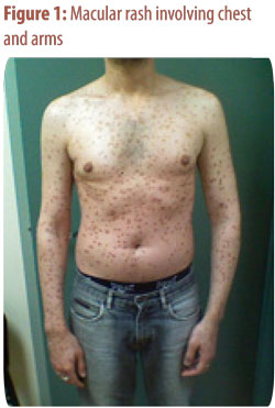
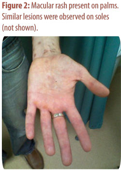
A 27-year-old MSM, presented to care with a rash. The rash appeared several weeks prior to presentation and involved the face, chest and back, arms and legs and was not accompanied by pruritus. He denied fever, chills, but complained of fatigue. No respiratory, gastrointestinal or urinary symptoms were present. He disclosed a diagnosis of HIV infection a year earlier, but has not kept his follow up appointments and was not receiving anti-retroviral medications or opportunistic infection prophylaxis. His most recent CD4 count was 109/mm3. He admitted sexual encounters with several male partners with inconsistent condom usage, and recalled a penile lesion that was present several weeks before the rash had appeared. The lesion has healed without specific therapy.

On physical examination: in no apparent distress, vital signs were within normal limits.

Notable finding on the examination included multiple small and non-tender anterior cervical, posterior cervical, axillary and inguinal lymph nodes. Genital examination revealed a healed lesion on the glans penis. A macular skin rash was widely distributed over face, trunk and extremities with several lesions on palms and soles (figure 1. and 2.)


1. What is your diagnosis?
2. Would you obtain a lumbar puncture?

Dysphagia among Older Adults

Dysphagia among Older Adults

Teaser: 

Amira Rana, MD, Medical Resident, Department Of Medicine, University of Toronto, ON.
Anselmo Mendez, BSc, BScN, Medical Student, Jagiellonian University, Medical College, Kraków, Poland.
Shabbir M.H. Alibhai, MD, MSc, FRCPC, Staff Physician, Department of Medicine, University Health Network; Assistant Professor, Departments of Medicine & Health Policy, Management, and Evaluation, University of Toronto, Toronto, ON.

Key words: myasthenia gravis, dysphagia, pyridostigmine, dysarthria, ptosis.

A Case of Methotrexate-Induced Pancytopenia

A Case of Methotrexate-Induced Pancytopenia

Teaser: 

David Wan, School of Medicine, University of Toronto, Toronto, ON.
Shabbir M.H. Alibhai, MD, MSc, FRCPC, Staff Physician, Department of Medicine, University Health Network, Toronto, ON; Assistant Professor, Departments of Medicine & Health Policy, Management, and Evaluation, University of Toronto.

Case Presentation
Mr. L was a 75-year-old man who presented in April 2007 with a one-month history of multiple discrete ulcerative lesions on his extremities, his trunk, and in his mouth. He had progressive weakness and severely restricted oral intake over the past two weeks due to pain from his oral ulcers...

Visual Hallucinations among Older Adults: The Charles Bonnet Syndrome

Visual Hallucinations among Older Adults: The Charles Bonnet Syndrome

Teaser: 

Nages Nagaratnam, MD, FRCP, FRACP, FRCPA, FACC, Consultant Physician in Geriatric Medicine, Department of Geriatric Medicine, Blacktown-Mt-Druitt Health, Blacktown, NSW, AUS.

A 73-year-old woman was seen in hospital troubled with visual hallucinations for over a year. They had increased in both frequency and intensity in the past few months, necessitating hospitalization. To her annoyance, the visual images took the form of people watching her through the glass windows. She less frequently saw animals. The hallucinations occurred both during the day and night but were worse as evening approached.

An Older Woman with Peripheral Arterial Disease -- May 2006

An Older Woman with Peripheral Arterial Disease -- May 2006

Teaser: 


From the Department of Medicine, Cardiology and Geriatrics Divisions, New York Medical College, Valhalla, NY, USA.
Wilbert S. Aronow, MD, CMD, Clinical Professor of Medicine, Divisions of Cardiology, Geriatrics, and Pulmonary/Critical Care Medicine;
Chief, Cardiology Clinic; Senior Associate Program Director and Research Mentor, Fellowship Programs, Department of Medicine, New York Medical College, Valhalla, NY, USA; Adjunct Professor of Geriatrics and Adult Development, Mount Sinai School of Medicine, New York City, NY, USA.

A 73-year-old woman saw a physician because of a three-month history of pain on walking after two blocks, which was relieved by rest. She had no other symptoms and was not on any medications.

Acute Low Back Pain:A Clinical Experience with Acupuncture

Acute Low Back Pain:A Clinical Experience with Acupuncture

Teaser: 


Sanjeev Rastogi, MD, CAc, Consulting Physician and Lecturer, Government Ayurvedic College and Hospital, Handia, Allahabad, India.
Rajieev Rastogi, MBSc, BNYS, Assistant Director (Naturopathy), Central Council for Research in Yoga and Naturopathy, New Delhi, India.

Acute low back pain (ALBP) is a common condition that results in huge economic losses in the form of treatment and absenteeism (direct monitary loss for incurring the treatment and indirect monitary loss resulting from absence). It responds well to conservative therapy, but it often takes a period of between one and three months before improvement is seen. Acupuncture has its effect in pain reduction in various musculoskeletal conditions including ALBP. As is observed in the present case, this technique can reduce the recovery period in these patients and, thus, can improve the net outcome.
Key words: acute low back pain, acupuncture, conservative management, recovery period, analgesia.