Holly Clark, Holly Clark is a freelance writer and works as a content manager for various international brands. When Holly is not researching and writing she loves nothing more than heading out into the country for some downtime. Holly is currently writing for Be in Health at https://www.beinhealth.com/
Caring for someone with dementia is never easy. In fact, many people often ask for support from other individuals who are also taking care of dementia patients. The first thing you should do is make some modifications in your home. People with dementia often see the world as a new and confusing place. Things can be scary every day. Therefore, it helps to adjust the environment to suit their needs and make them feel as comfortable as possible. Here is a guide on how individuals can create a danger-free home for those with dementia.
Ask for Guidance from Local Aging Agency
Often, these professionals have experience in taking care of people with Alzheimer's or other diseases related to dementia. They can recommend certified aging experts to come inspect your house and advise accordingly on safety issues in your home. It, therefore, helps to give them a call before modifying your house.
Inspect Areas of Your House That May be Unsafe
Check out areas in your house that you suspect may compromise the safety of your loved one. And when doing that, it helps to inspect them from the patient's point of view. So you have to remember that this condition affects one's balance, cognitive abilities, memory as well as perception. According to Jane Byrne, Project Coordinator at FirstCare nursing home Wicklow, "Someone with this condition has a difficult time interpreting and remembering information as well as making sound decisions." It, therefore, helps to modify your house in a way that will give them an easy time. And when doing that, you also should be careful. Redecorating or redesigning your home and other significant changes can be unsettling. On the other hand, simple moves like furniture rearrangement can also be alarming. Therefore, be cautious and give them time to adjust.
Follow the AARP Checklist for Home Safety
For those who don't have any idea on where to start, having this list will be useful. It outlines all the safety measures caregivers should take in order to make a home danger-free for loved ones with dementia. This list will serve as a guide on how to create a safe environment. Some of the things homeowners are advised to do, include:
Decluttering the walkway of any substance. Even if there is ice, snow, or debris, individuals should make sure it is removed. It's essential that you move bikes, chalks, lawn ornaments, or jump ropes to other rooms.
Marking Step Edges Using Neon Glow in the Dark Tape
Providing sufficient lighting, both indoors and outdoors. Sometimes people with dementia may perceive shadows as demons or burglars. Placing bright outdoor lighting, therefore, helps reduce falls, fear, as well as anxiety.
Repairing uneven bricks or cracked pavements as they serve as tripping points.
Designate a Danger Zone
Dementia people forget how things are used quickly. They can even drink wiper juice or touch a hot grill with bare hands. Therefore, individuals should convert one room, either the garage or outdoor shed, into storing substances such as sharp knives, as well as cutters, bleach, washing products, paint, insecticides, and other substances that might be confused. It also helps to have a combination of locks on rooms that contain hazardous items.
Creating a safe home for seniors with dementia is about identifying areas in your house that may pose dangers and modifying them so that they're safe. It also helps that individuals do a pantry patrol regularly since patients may eat spoiled or moldy foods.


 For the 6 months that I have not been able to drive because of medical reasons I have become a regular user of Uber. I find Uber more flexible and convenient than standard taxis. As regular users of Uber services know, many of the drivers are originally from elsewhere, by which I mean overseas and came as immigrants or refugees.
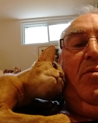
 The driver and I talked about the wonderment of cats and how they are part of Istanbul life. To observe the seven cats and their litters featured in the movie was a real joy, with the film starting with a picture of kittens waiting for their mamma to return with breakfast—their tiny heads peering through the railing surrounding the birth site. The clips of the cat who ventures to the fish market every morning where the local fishmonger prepares the remnants of the carved fish for the dependable visitor as if it were a guest coming for dinner. Among the most moving scenes were that of people whose lives were either impacted or even saved by their feline companion who through the special sense that cats have of responding to human need.