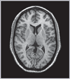
 Are structural changes in the brain a normal byproduct of the aging process? This complex question can be addressed by separately evaluating the contribution of four commonly observed changes--cortical atrophy (increased sulcal cerebrospinal fluid), central atrophy (ventricular enlargement), deep white matter hyperintensities (DWMHs) and periventricular hyperintensities (PVHs)--all of which can be categorized as forms of subclinical structural brain disease (SSBD). Although these structural changes predominate in demented and depressed patients, they also have been implicated as features of normal aging. However, the mechanism by which these changes contribute to cognitive decline has yet to be elucidated.
In a cross-sectional study, Cook and colleagues examined the relationship between structural changes, age, cognition and the connectivity between brain regions. A combination of magnetic resonance imaging (MRI), neuropsychological testing and quantitative electroencephalographics (QEEG) were used to asses SSBD volumes, cognition levels and functional connectivity, respectively.
A series of hypotheses were made, predicting a correlation between increasing age and increasing SSBD volume, between increasing SSBD volume and decreasing cognitive performance and between increasing SSBD volume and increased disconnectivity between brain regions. In total, the proposal sought to show that brain region connectivity mediates the effects of increased SSBD volume on reduced cognitive function.
The study involved 43 healthy subjects aged 60-93 years. The brains of the recruits were scanned using MRI to measure the volumes of specific tissue and fluid types. QEEG coherence was calculated separately in the prerolandic and postrolandic regions to evaluate the degree of connectivity between brain regions via well-recognized neuroanatomical pathways. The Trail-Making Tests (Trail A to measure attention and processing speed; Trail B to measure sequencing ability) were among many of the neuropsychological tests employed.
A linear relationship was established between age and central and cortical atrophy and between age and PVHs, but was not seen between age and DWMHs. While the general trend was towards greater SSBD volume in older subjects, many subjects older than 75 in fact displayed smaller SSBD volumes than their younger counterparts. Furthermore, a greater variability in SSBD volume was observed with increasing age. Thus, this result on its own is somewhat uninformative.
As hypothesized, increased SSBD volume was found to correlate with an overall decrease in cognitive function. This result was most strongly evinced in Trails B, a test involving a series of processing steps, suggesting that structural changes in the aging brain may make multi-stepped processes more difficult, while exerting a less noticeable impact on simple tasks. QEEG recordings demonstrated that increasing PVH, DWMHs and ventricular cerebrospinal fluid (vCSF) were associated with lower coherence levels in both prerolandic and postrolandic networks.
Finally, a path analysis model demonstrated a mediation role for connectivity disruption in the effect of white matter disease on cognition, but failed to confirm the same association for atrophy.
Overall, these results support the notion that increased SSBD volume negatively affects cognition via neuronal connectivity disruption in normal aging. To further bolster this theory, future research might explore the results from task-activated QEEG recordings, rather than the resting-state EEGs that were used in this study.
Source
- Cook IA, Leuchter AF, Morgan ML, et al. Cognitive and physiologic correlates of subclinical structural brain disease in elderly healthy control subjects. Arch Neurol 2002;59:1612-20.

 Are structural changes in the brain a normal byproduct of the aging process? This complex question can be addressed by separately evaluating the contribution of four commonly observed changes--cortical atrophy (increased sulcal cerebrospinal fluid), central atrophy (ventricular enlargement), deep white matter hyperintensities (DWMHs) and periventricular hyperintensities (PVHs)--all of which can be categorized as forms of subclinical structural brain disease (SSBD). Although these structural changes predominate in demented and depressed patients, they also have been implicated as features of normal aging. However, the mechanism by which these changes contribute to cognitive decline has yet to be elucidated.