Advertisement

Advertisement

colonoscopy

Things that fascinate me about radiology

0

No applauses yet

I was a family physician for 7 years before becoming a radiologist. There are some things I miss about family practice. I miss the longitudinal relationship that I often had with multiple generations of family members.

There are also some things that fascinate me about radiology. For instance, improving computer and imaging technology has translated into imaging developments that can be used to benefit patients. Just a few years ago, we wouldn’t have thought we could adequately screen the colon for colon cancer and pre-cancerous lesions such as polyps. Now, in patients who cannot have a colonoscopy, we routinely perform CT colonoscopy (CTC), giving an alternative means of screening and diagnosis in these patients.

An article in the New England Journal of Medicine suggests that CTC could be used for primary screening, however, we mainly use it for patients who have failed colonoscopy – often because of a redundant sigmoid colon which cannot be navigated by the scope. We can even do the study the same day, as the patient has already undergone a bowel preparation. We do give them contrast to tag any residual fluid and stool in the colon to be able to differentiate it from colonic pathology.

We perform CT prone and supine to allow us to exam all the walls of the colon without any overlying fluid. Then we use computer software to generate 3 dimensional images of the colon that we can “fly through” to assess for mucosal lesions. Of course, the CT images are also examined for any extra- mucosal findings. I have diagnosed an unsuspected renal cell carcinoma on a patient being screened for colon cancer.

Below is a picture from my practice. It shows the colon distended with carbon dioxide – we use a small rectal tube and a regulated pump to inflate the colon. The technique readily shows the “napkin-ring” constricting lesion in the cecum. The 3D images show the lesion as it would be seen by the scope!

I would love to hear how imaging has affected your practice, both positive and negative.

Reference:
CT Colonography versus Colonoscopy for the Detection of Advanced Neoplasia
David H. Kim, M.D., Perry J. Pickhardt, M.D., Andrew J. Taylor, M.D., Winifred K. Leung, M.D., Thomas C. Winter, M.D., J. Louis Hinshaw, M.D., Deepak V. Gopal, M.D., Mark Reichelderfer, M.D., Richard H. Hsu, M.D., and Patrick R. Pfau, M.D.
N Engl J Med 2007; 357:1403-1412; October 4, 2007
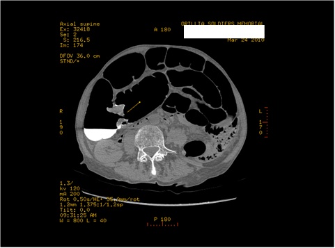


Axial view with the “Napkin-ring” mass seen in the region of the cecum-ascending colon.
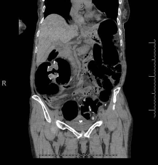


Coronal Image showing the lesion.
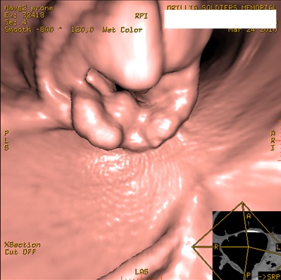


3D image showing the lesion almost identical to how it would appear on Colonoscopy, had this patient been able to have colonoscopy.

Colorectal Cancer: A Disease with a Promising Future

Colorectal Cancer: A Disease with a Promising Future

Teaser: 


The accredited CME learning activity based on this article is offered under the auspices of the CE department of the University of Toronto. Participating physicians are entitled to one (1) MAINPRO-M1 credit by completing this program, found online at www.geriatricsandaging.ca/cme.htm

J. A. Maroun, MD, Medical Oncologist, Integrated Cancer Program, The Ottawa Hospital; Professor of Medicine, University of Ottawa, Ottawa, ON.

Colorectal cancer is one of the most common cancers in Canada. In the last decade, there has been significant progress in the management of this disease. Improved understanding of the pathophysiology of colorectal cancer has resulted in the development of more prevention and screening strategies. Adjuvant therapy in high-risk patients has led to an increase in cure rates. For years, 5-fluorouracil was the only drug available for metastatic disease; now, new and effective drugs have been developed, with opportunities for effective second- and third-line therapies as well as new combinations. This has led to an increase in the median survival of patients from six months to over 20 months. Ongoing research with new agents—in particular, biologically targeted drugs—will undoubtedly lead to further improvement in the outcome of this disease.

Key words: colorectal cancer, 5-fluorouracil, colonoscopy, chemotherapy, radiation.

Diverticular Disease of the Colon: Review and Update

Diverticular Disease of the Colon: Review and Update

Teaser: 

Christopher N. Andrews, MD, Gastroenterology Fellow, Faculty of Medicine, University of Calgary, Calgary, AB.
Eldon A. Shaffer, MD, FRCPC, Professor of Medicine, Division of Gastroenterology, Faculty of Medicine, University of Calgary, Calgary, AB.

Introduction
Diverticular disease of the colon (or diverticulosis) is an anatomical description of saccular outpouchings of mucosa through the wall of the colon. It is very common in the Western world, and its prevalence is rising. This paper will briefly review the epidemiology and pathophysiology of diverticular disease, followed by a focus on the diagnosis and management of the two most common complications of the disease: diverticulitis and diverticular bleeding.

Epidemiology
The true prevalence of diverticulosis is unknown, but autopsy reports suggest that up to half of patients over 60 years are affected.1 The frequency increases with age and is much higher in developed societies in which fibre intake is lower. In the Western world, the most commonly affected site in the colon is the sigmoid colon, sometimes with more proximal involvement.2 However, in Asian countries diverticulae tend to be right-sided (in the ascending colon) and fewer in number. The reason for this difference is unknown.

Pathophysiology
The colon is made up of circumferential and longitudinal (taenia coli) muscle layers, which act in unison to propel stool towards the rectum.

Colonoscopy Free?

Colonoscopy Free?

Teaser: 

Apparently, not yet. However, promising results from a study in the New England Journal of Medicine capitalize on our understanding of the genetic basis behind colon cancer. Colon cancer arises when four or five genes become mutated. In more than 90% of cases, it is mutations in the adenomatous polyposis coli (APC) gene that initiate colorectal tumours. The researchers investigated whether it was possible to detect APC mutations in fecal DNA using a new developed method known as digital protein truncation, a technique that could identify mutations in a sensitive and specific manner. Stool samples were collected from 28 patients with nonmetastatic colorectal cancers, 18 patients with adenomas that were at least 1 cm in diameter and 28 control patients. The test managed to identify mutations in 26 of the 46 patients with neoplasia (57%), and in none of the control patients.

Although successful in preventing false positives, which is a common occurrence with the current method of checking the stool for blood, the fact that the method only identified cancers in 50% of the patients suggests that this is only the first step in a long process. However, the researchers did overcome the somewhat unenviable task of sorting through the stool samples and managed to identify APC genes in each of the 74 stool samples.

Further research is underway, and the group predicts that an accurate and practical version of the test may be available within the next five years.

Source

  1. Traverso GT, Shuber A, Levin B et al. Detection of APC mutations in fecal DNA from patients with colorectal tumours. NEMJ 2002;346:311-320.

Virtual Colonoscopy--Non-invasive Procedures Holds Many Advantages for the Patient

Virtual Colonoscopy--Non-invasive Procedures Holds Many Advantages for the Patient

Teaser: 

Anna Liachenko, BSc, MSc
Managing Editor, Geriatrics & Aging

Despite the fact that colon cancer is preventable, over 16,000 elderly Canadians were diagnosed with the disease in 1999. One out of every three patients diagnosed with the disease, died. Prevention of colon cancer requires discovery and removal of the precursor polyp at an early stage. However, elderly patients, who are at risk of developing colon cancer are largely underscreened. The reasons range from high invasiveness of current techniques and, thus, poor patient acceptance, to inability of the current methods to efficiently detect small polyps. Fortunately, a new non-invasive and highly efficient technique--virtual colonoscopy--has recently been introduced. This article will describe the technique and compare this technique with the traditional methods of colonoscopy and barium enema.

David J. Vining and associates, at the Bowman Gray School of Medicine first described virtual colonoscopy in 1994. It is based on analysis of two sets of axial images obtained from thin-section helical computed tomography (CT) scans of the abdomen and pelvis. The procedure starts with cleansing the patient's bowel (using a standard barium enema or colonoscopy bowel preparation) followed by colonic insufflation with room air or carbon dioxide. Then, the abdomen and pelvis are scanned with the patient holding their breath for the first 15 to 20 seconds (to cover the upper abdomen) and gently respiring for the remainder of the scan.