Radiologist, Orillia Soldiers' Memorial Hospital, Adjunct Clinical Lecturer, Department of Family and Community Medicine and Department of Medical Imaging, University of Toronto, Toronto, ON.
Abstract: Prostate cancer is a common cancer in men worldwide, and early detection is key to improved patient outcomes. Diagnosis typically involves a combination of clinical examination, prostate-specific antigen blood testing, and imaging studies. Radiology plays an important role, aiding in treatment planning, confirming the diagnosis by directing biopsy, staging the patient, and following treatment course. Imaging modalities for prostate cancer diagnosis include ultrasound, CT, nuclear medicine, and MRI. While MRI is the most sensitive imaging modality, ultrasound is still the preferred modality for measuring the prostate volume. Prostate-specific membrane antigen PET imaging has shown to have superior sensitivity and specificity compared to conventional imaging modalities in the detection of prostate cancer, especially in the context of low PSA. Clinical pearls include performing ultrasound-guided biopsy under local anesthesia to improve patient comfort, and the use of fusion MRI and ultrasound images to facilitate MRI/TRUS fusion-guided biopsy.
You can take quizzes without subscribing; however, your results will not be stored. Subscribers will have access to their quiz results for future reference.
Radiology plays a crucial role in prostate cancer diagnosis, aiding in treatment planning, confirming the diagnosis, and directing biopsy.
Imaging modalities for prostate cancer diagnosis include ultrasound, CT, nuclear medicine, and MRI.
MRI is the most sensitive conventional imaging modality for detecting prostate cancer.
Prostate-specific membrane antigen PET imaging has been shown to have superior sensitivity and specificity compared to conventional imaging modalities in the detection of prostate cancer, especially in context of low PSA.
Ultrasound is still the preferred modality for measuring the prostate volume.
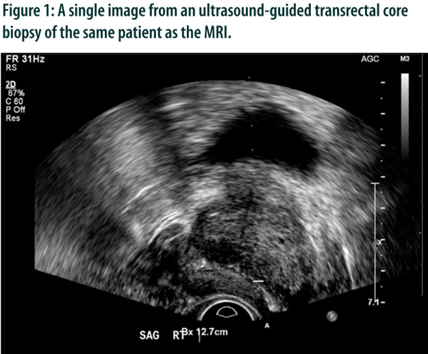
Ultrasound-guided biopsy is a minimally-invasive procedure that involves inserting a needle through the rectum via an ultrasound probe guide and into the prostate gland. It is performed under local anesthesia and patients are discharged the same day after a short period of observation in the radiology department.
MRI and ultrasound images can be fused to facilitate MRI/TRUS fusion-guided biopsy, which improves the accuracy of the biopsy procedure.
The use of antibiotic prophylaxis before ultrasound-guided biopsy decreases the risk of infection to approximately 1 in 100 patients.
To have access to full article that these tools were developed for, please subscribe. The cost to subscribe is $80 USD per year and you will gain full access to all the premium content on www.healthplexus.net, an educational portal, that hosts 1000s of clinical reviews, case studies, educational visual aids and more as well as within the mobile app.