1Michael G. DeGroote School of Medicine, McMaster University, Hamilton, Ontario. 2Clinical Associate Professor, Department of Pediatrics, Associate Member, Department of Dermatology and Skin Sciences, University of British Columbia, Vancouver, BC.
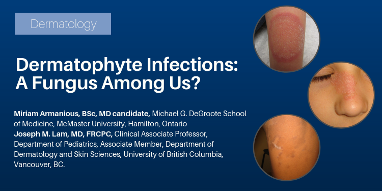
Abstract: Superficial fungal infections are a common occurrence in adults and children alike. Dermatophytes are the primary cause of these infections, which generally present as erythematous, scaling, annular lesions. Also referred to as "tinea", these infections are classified based on where they are found on the body, as different locations can have slightly different presentations and treatment requirements. This article provides an overview of these various presentations of dermatophyte infections and their risk factors, as well as recommended therapies.
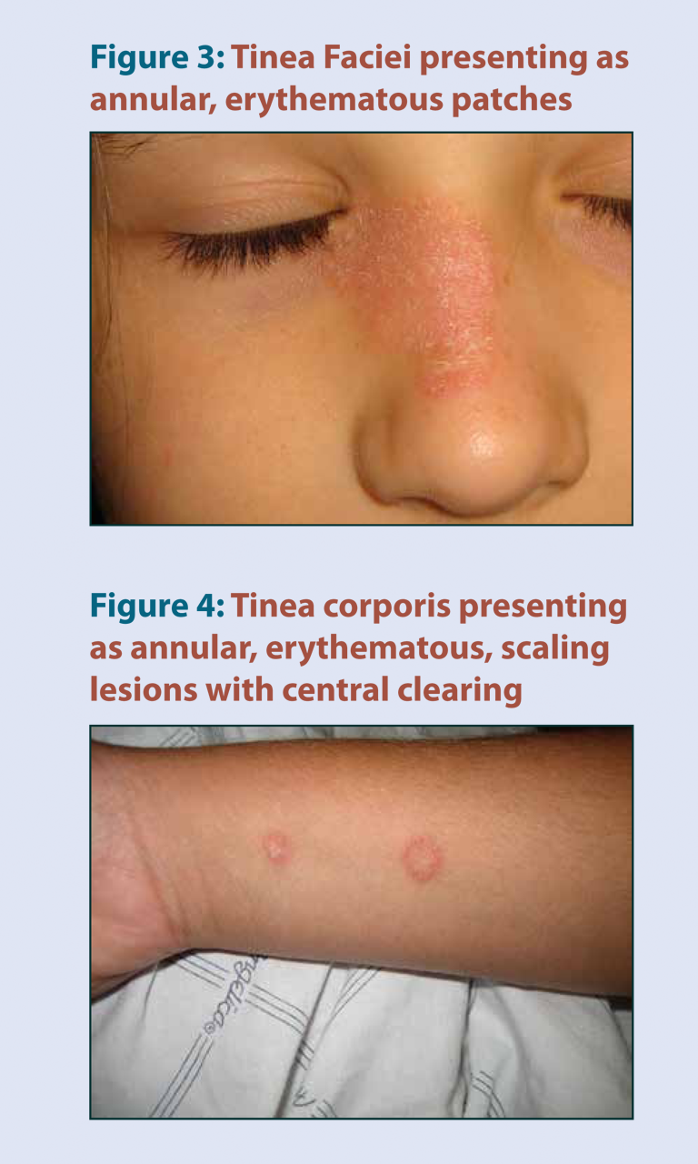
Dermatophyte infections, also known as tinea, are very common fungal infections in humans. They occur on the superficial skin, hair, and nails, and can present in many different locations on the body.
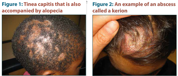
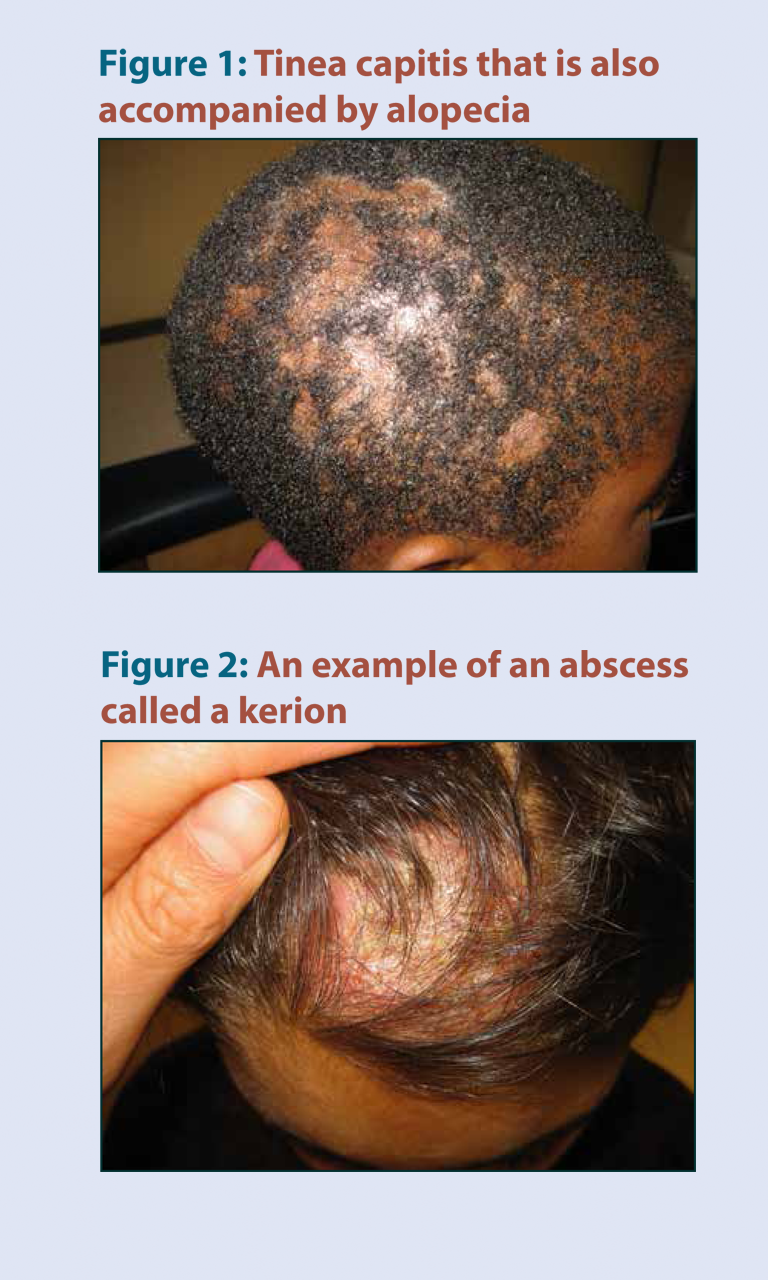
Tinea captis is most common in children and can cause hair loss or abscess formation.
When tinea infections are treated with topical corticosteroids, they become harder to detect and are referred to as tinea incognito.
Tinea infections are common, but should be confirmed with KOH microscopy and/or culture from a skin scraping, nail clipping,
or hair sample.
Tinea capitis can be mistaken for eczema or seborrheic dermatitis
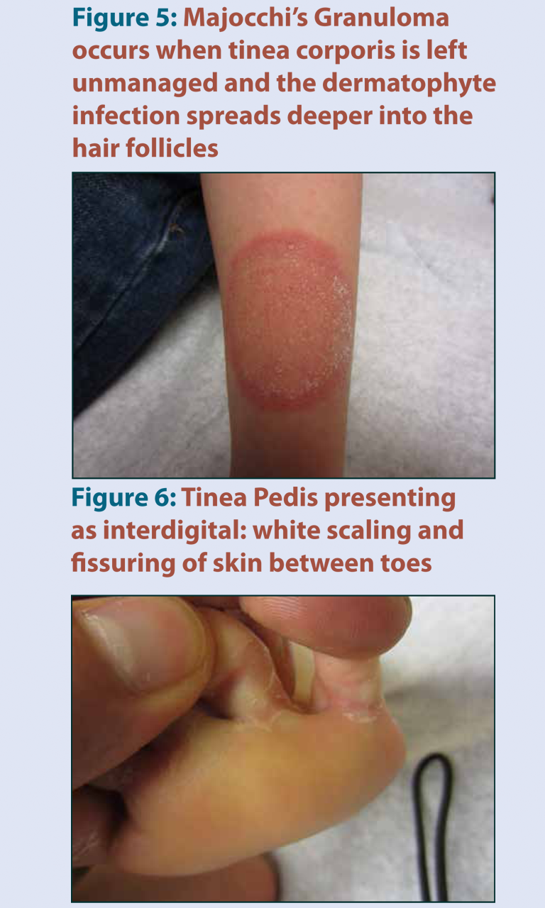
Check patients who have tinea infection for tinea pedis, since this is a common source of infection for sites on the rest of the body
Treatment for dermatophyte infections can include oral antifungal agents such as terbinafine or grise-ofulvin in a weightdependent
dose, or topical antifungal agents. Systemic agents are generally re-served for presentations that penetrate hair follicles and nails, or those that are refractory to topical treatment.
To have access to full article that these tools were developed for, please subscribe. The cost to subscribe is $80 USD per year and you will gain full access to all the premium content on www.healthplexus.net, an educational portal, that hosts 1000s of clinical reviews, case studies, educational visual aids and more as well as within the mobile app.