Advertisement

Advertisement

bone mineral density

Bone Mineral Density: What Is Its Relationship to Heart Disease?

Bone Mineral Density: What Is Its Relationship to Heart Disease?

Teaser: 

Wilbert S. Aronow, MD, FACC, FAHA, AGSF, FCCP, Divisions of Cardiology, Geriatrics, and Pulmonary/Critical Care, Department of Medicine, New York Medical College, Valhalla, NY, USA.

Low bone mineral density (BMD) is associated with obstructive coronary artery disease (CAD); this article reviews several recent studies that have demonstrated the association. In one study, for every 1-unit reduction in femoral neck T score, a 0.23 minute decrease in treadmill exercise duration was found after values were adjusted for age and other patient characteristics (95% confidence interval [CI], 0.11–0.35, p<0.001). For every 1-unit reduction in femoral neck T score, there was a 22% increased risk of myocardial ischemia after values were adjusted for age and other patient characteristics (95% CI, 1.06–1.41, p = 0.004). Overall after adjustments, patients with a low BMD who were referred for exercise echocardiographic stress testing had a 43% greater risk of myocardial ischemia than did patients with normal BMD referred for exercise echocardiographic stress testing (95% CI, 1.06–1.94, p = 0.02). Reduced physical activity may contribute to both low BMD and CAD through the development of atherosclerotic vascular disease.
In a second study, stress test-induced myocardial ischemia developed in 95 of 254 patients (37%) with osteoporosis, in 81 of 260 patients (31%) with osteopenia, and in 62 of 251 patients (25%) with normal BMD (p= 0.009) (p= 0.002 comparing osteoporosis with normal BMD; p=0.007 comparing osteoporosis or osteopenia with normal BMD). Patients with osteoporosis or osteopenia had a 1.7 times higher chance of stress test-induced myocardial ischemia than those with normal BMD after controlling the confounding effects of systemic hypertension, diabetes mellitus, body mass index, and age.
Key words: osteoporosis, osteopenia, bone mineral density, coronary artery disease, myocardial ischemia.

Viser le ligand RANK augmente la densité minérale osseuse chez les femmes ménopausées : résultats d’essais de phase 3

Viser le ligand RANK augmente la densité minérale osseuse chez les femmes ménopausées : résultats d’essais de phase 3

Teaser: 

Le rôle du mécanisme RANK/RANKL/OPG dans la perte osseuse : nouvelles perspectives

Viser le ligand RANK augmente la densité minérale osseuse chez les femmes ménopausées : résultats d’essais de phase 3

Conférencière : Alexandra Papaioannou, M.D., M.Sc., FRCPC, Professeure, Directrice du département de médecine, Université McMaster, Gériatre, Hamilton Health Sciences Centre, Hamilton, ON.

La Dre Alexandra Papaioannou a passé en revue les résultats d’essais de phase 3 sur le dénosumab qu’elle a décrit comme un « nouveau produit passionnant » qui fait l’objet de recherches. Tel que l’a expliqué le Dr Robert Josse, le dénosumab est un anticorps monoclonal (IgG2) entièrement humain qui se lie avec haute affinité et spécificité au ligand RANK (receptor activator of nuclear factor kappa B) (RANKL) humain. RANKL est un médiateur essentiel de l’activité des ostéoclastes.1-3 La Dre Papaioannou a noté que les essais cli-niques n’ont jusqu’ici pas découvert d’anticorps neutralisants1,3 et que les effets du dénosumab sur la résorption osseuse semblent être réversibles.3

Le dénosumab qui réduit les marqueurs du renouvellement des cellules osseuses est administré par injection sous-cutanée tous les 6 mois, ce qui est avantageux chez une population plus âgée. Il a une demi-vie de ~25 à 46 jours.4

La Dre Papaioannou a passé en revue quatre études de phase 3 sur le dénosumab en présence d’ostéopénie ou d’ostéoporose postménopausique.

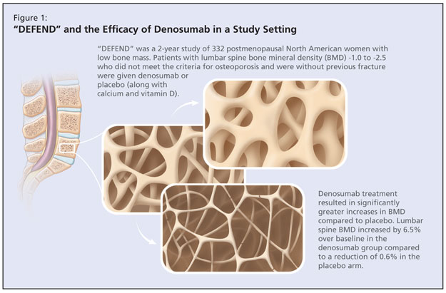
L’étude DEFEND était une étude randomisée à double insu, contrôlée par placebo qui visait à déterminer si un traitement par dénosumab pourrait prévenir la perte osseuse de la colonne lombaire chez les femmes ménopausées souffrant d’ostéopénie (Figure 1).4


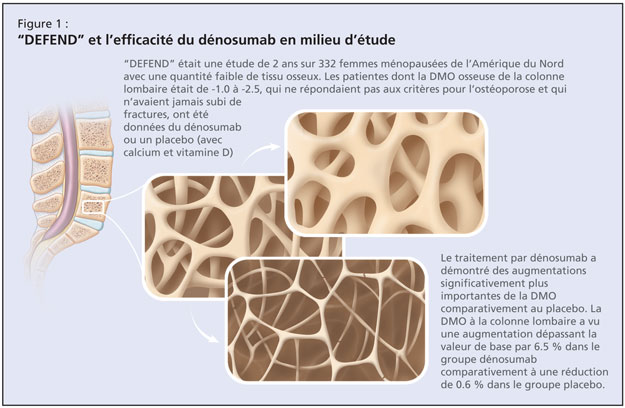
Cette étude de 2 ans sur 332 femmes ménopausées de l’Amérique du Nord chez lesquelles la quantité de tissu osseux était faible a étudié les effets du dénosumab ou d’un placébo chez les patientes dont la densité minérale osseuse (DMO) à la colonne lombaire était de –1.0 à –2.5, qui ne répondaient pas aux critères pour l’ostéoporose et qui n’avaient jamais subi de fractures. L’âge moyen était de 58 ans, ce qui constitue une population plutôt jeune, comme l’a noté la Dre Papaioannou. La valeur de base moyenne du score-T de la DMO à la colonne lombaire des participantes était de –1.61. Les patientes ont reçu par randomisation, soit 60 mg de dénosumab, soit un placebo pendant une période de 2 ans. Toutes les patientes ont reçu chaque jour, ≥1000 mg de calcium et ≥400 UI de vitamine D. Le paramètre primaire était la variation en pourcentage de la DMO à la colonne lombaire à 24 mois. Les résultats publiés ont démontré qu’un traitement par dénosumab entraîne des augmentations de la DMO significativement plus importantes pour tous les endroits mesurés, comparativement au placebo (P < 0.05; 95 % intervalle de confiance [IC]). La DMO à la colonne lombaire a vu une augmentation dépassant la valeur de base par 6.5 % dans le groupe dénosumab, comparativement à une baisse de 0.6 % dans le groupe placebo. Des effets secondaires se sont produits dans chaque groupe, les plus communs étant l’arthralgie, la rhinopharyngite et les maux de dos. Il y avait une légère tendance de sérieux évènements indésirables dans le groupe dénosumab.

L’étude DECIDE était une étude randomisée à double insu, comparative avec traitement de référence, qui a évalué les effets du dénosumab comparativement à l’alendronate sur la variation en pourcen-tage de la DMO à la hanche totale à 12 mois, sur 1189 femmes ménopausées.5 Ceci était une étude de non-infériorité sur des femmes ménopausées (≥ 12 mois) avec une faible quantité de tissu osseux et un score-T ≤ –2.0 à la colonne lombaire et à la hanche totale. L’âge moyen des participantes était de 64 ans; ~24 % avaient préalablement reçu une thérapie médicale pour l’ostéoporose et jusqu’à 50 % avaient déjà subi une fracture. Les patientes ont reçu par randomisation des injections de dénosumab (60 mg tous les 6 mois) en plus d’un placebo oral chaque semaine ou de l’alendronate orale chaque semaine (70 mg) en plus d’injections sous-cutanées du placebo tous les 6 mois. Le paramètre primaire était un changement de la DMO à la hanche totale après une période de 1 an. Les résultats publiés ont démontré qu’un traitement par dénosumab entraîne des augmentations significativement plus importantes du changement en pourcentage à partir de la valeur de base de la DMO pour tous les endroits du squelette mesurés, comparativement à l’alendronate. Des augmentations statistiquement significatives de la DMO à la hanche totale ont été observées chez le groupe dénosumab comparativement à ceux traités par alendronate (3.5 % vs 2.5 %; P < 0.0001). Le taux d’abandon était semblable pour chaque groupe et la fréquence et le type d’évènements indési-rables et de sérieux évènements indésirables étaient comparables.

La Dre Papaioannou a ensuite exposé les détails de l’étude STAND, une étude de phase 3 randomisée à double insu, comparative avec traitement de référence, sur des femmes préalablement traitées par alendronate.6 Elle a décrit cette étude comme étant d’une utilité particulière étant donné la fréquence du scénario du patient suivant un régime d’alendronate qui cherche à changer de traitement. L’étude a évalué les effets d’une transition au dénosumab sur les changements de la DMO et des marqueurs biochimiques du renouvellement des cellules osseuses, et sur la sécurité et la tolérabilité, comparativement à la continuation d’un traitement par alendronate. Les participantes étaient des femmes ménopausées (âge moyen de 68 ans) avec une DMO faible, préalablement traitées avec 70 mg d’alendronate une fois par semaine ou l’équivalent pour ≥6 mois. Leurs mesures de la DMO corres-pondaient à un score-T ≤ –2.0 et ≥ –4.0 à la hanche totale. Les patientes ont reçu par randomisation des injections de dénosumab (60 mg tous les 6 mois) ou de l’alendronate orale (70 mg). Le paramètre primaire était la variation en pourcentage à partir de la valeur de base de la DMO à la hanche totale à 12 mois. Les auteurs ont signalé que la DMO à la hanche totale a vu au douzième mois, une augmentation de 1.90 % à partir de la valeur de base, chez des sujets faisant la transition au dénosumab, comparativement à une augmentation de 1.05 % à partir de la valeur de base chez des sujets continuant la thérapie par alendronate (P < 0.0001). Les évènements indésirables, les sérieux événements indésirables d’infections et les néoplasmes étaient comparables entre les deux groupes expérimentaux.

Finalement, la Dre Papaioannou a exposé les détails de l’étude FREEDOM, une étude randomisée à double insu, contrôlée par placebo qui visait à déterminer si un traitement par dénosumab réduit le nombre de femmes ménopausées qui souffrent de nouvelles fractures vertébrales.7 L’étude en cours comporte 7868 femmes ménopausées (-4.0 < DMO score-T < –2.5) âgées de 60 à 90 ans et exclue les individus avec de sévères fractures ou celles plus nombreuses que deux. Les patientes reçoivent par randomisation des injections de dénosumab (60 mg tous les 6 mois) ou un placebo. Les paramètres primaires comprennent la fréquence de nouvelles fractures vertébrales en plus du profil de sécurité et de tolérabilité du dénosumab.

Références :

  1. Bekker PJ, Holloway DL, Rasmussen AS, et al. A single-dose placebo-controlled study of AMG 162, a fully human monoclonal antibody to RANKL, in postmenopausal women. J Bone Miner Res 2004;19:1059–1066.
  2. Elliott R, Kostenuik P, Chen C, et al. Denosumab is a selective inhibitor of human receptor activator of NF-Kb ligand (RANKL) that blocks osteoclast formation and function. Osteoporos Int 2007;18:S54. Abstract P149.
  3. McClung MR, et al. Denosumab in post-menopausal women with low bone mineral density. New Engl J Med 2006;354:821–31.
  4. Bone HG, Bolognese MA, Yuen CK, et al. Effects of denosumab on bone mineral density and bone turnover in postmenopausal women. J Clin Endocrinol Metab 2008;93:2149–57.
  5. Brown JP, Prince RL, Deal C, et al. Comparison of the effect of denosumab and alendronate on BMD and biochemical markers of bone turnover in postmenopausal women with low bone mass: a randomized, blinded, phase 3 trial. J Bone Miner Res 2009;24:153–61.
  6. Kendler DL, Benhamou CL, Brown JP et al. Effects of denosumab vs alendronate on bone mineral density (BMD), bone turnover markers (BTM), and safety in women previously treated with alendronate. J Bone Miner Res 2008;23(suppl 1):S473. Abstract M395 and poster.
  7. Cummings SR, McClung MR, Christiansen C, et al. A phase III study of the effects of denosumab on vertebral, nonvertebral, and hip fracture in women with osteoporosis: Results from the FREEDOM trial. J Bone Miner Res 2008;23(suppl 1):S80. Abstract 1286 and oral presentation.

Cette présentation a été appuyée par une subvention éducationnelle sans restrictions accordée par Amgen Canada.

Targeting RANK Ligand Increases Bone Mineral Density||in Postmenopausal Women: Results from Phase 3 Trials

Targeting RANK Ligand Increases Bone Mineral Density||in Postmenopausal Women: Results from Phase 3 Trials

Teaser: 


The Role of RANK/RANKL/OPG Pathway in Bone Loss: New Insights

Targeting RANK Ligand Increases Bone Mineral Density in Postmenopausal Women: Results from Phase 3 Trials

Speaker: Alexandra Papaioannou, MD, MSc, FRCPC, Professor, Department of Medicine Director, Division of Geriatric Medicine, McMaster University, Geriatrician, Hamilton Health Sciences Centre, Hamilton, ON.

Dr. Alexandra Papaioannou reviewed the results of Phase 3 trials of denosumab, which she described “an exciting new compound” under investigation. As Dr. Robert Josse had explained, denosumab is a fully human monoclonal antibody (IgG2) that binds with high affinity and specificity to human RANK (receptor activator of nuclear factor kappa B) ligand (RANKL). RANKL is an essential mediator of osteoclast activity.1-3 Dr. Papaioannou noted that no neutralizing antibodies have been detected in clinical trials to date,1,3 and that the effects of denosumab on bone resorption appear reversible.3

Denosumab, which reduces bone turnover markers, is delivered via subcutaneous injection every 6 months, an advantage in an older patient population. It has a mean half-life of ~25–46 days.4

Dr. Papaioannou reviewed four Phase 3 studies of denosumab in post-menopausal osteopenia and/or osteoporosis.

The DEFEND study was a randomized, double-blind, placebo-controlled study that aimed to determine whether denosumab treatment could prevent lumbar spine bone loss in menopausal women with osteopenia (Figure 1).4 This 2-year study of 332 postmenopausal North American women with low bone mass investigated denosumab or placebo in patients with lumbar spine bone mineral density (BMD) -1.0 to -2.5 who did not meet the criteria for osteoporosis and were without previous fracture. Mean age was 58 years, which Dr. Papaioannou noted to be a younger population. The baseline mean lumbar spine BMD T-score of participants was -1.61. Participants were randomized to either 60 mg denosumab or placebo for 2 years. All patients received ≥1,000 mg of calcium and ≥400 IU vitamin D daily. The primary endpoint was percent change in lumbar spine BMD at 24 months. According to published results, denosumab treatment resulted in significantly greater increases in BMD at all measured sites compared to placebo (P < 0.05; 95% confidence interval [CI]). Lumbar spine BMD increased by 6.5% over baseline in the denosumab group compared to a reduction of 0.6% in the placebo arm. Adverse events occurred in both arms, the most commonly reported of which were arthralgia, nasopharyngitis, and back pain. There was a slight trend in the denosumab arm for serious adverse events.


The DECIDE study was a randomized, double-blind, active-controlled study that evaluated the effect of denosumab compared to alendronate on percent change in BMD in the total hip at 12 months in 1,189 postmenopausal women.5 This was a noninferiority study of postmenopausal (≥ 12 months) women with low bone mass and a T-score ≤ –2.0 at lumbar spine or total hip. Participants were a mean age of 64 years; ~24% had previously used medical therapy for osteoporosis and up to 50% had prior fracture. Patients were randomized to denosumab injections (60 mg every 6 months) plus oral placebo weekly or oral alendronate weekly (70 mg) plus subcutaneous placebo injections every 6 months. The primary endpoint was change in BMD at total hip after 1 year. According to the published results, denosumab treatment resulted in significantly greater increases in the percent change from baseline in BMD at all skeletal sites measured compared with alendronate. Statistically significant increases in BMD at the total hip were observed for the denosumab group compared with those treated with alendronate (3.5% vs 2.5%; P < 0.0001). Dropout rates were similar in both arms, and the incidence and type of adverse and serious adverse events were balanced.

Dr. Papaioannou next detailed the STAND study, a randomized, double-blind, active-controlled Phase 3 study of women previously treated with alendronate.6 She described this study as having particular utility given the common scenario of patients on a regime of alendronate who seek to switch treatment. The study evaluated the effects of transitioning to denosumab on changes in BMD and biochemical markers of bone turnover, and on safety and tolerability compared with continuation of alendronate therapy. Participants were postmenopausal women (mean age 68 years) with low BMD previously treated with alendronate 70 mg once weekly or equivalent for ≥6 months. Their BMD measurements corresponded to a T-score ≤ –2.0 and ≥ –4.0 at the total hip. Patients were randomized to denosumab injections (60 mg every 6 months) or oral alendronate weekly (70 mg). The primary endpoint was the percent change from baseline in total hip BMD at 12 months. The authors reported that BMD at the total hip increased by 1.90% from baseline at month 12 in subjects transitioning to denosumab compared with a 1.05% increase from baseline in subjects continuing on alendronate therapy (P < 0.0001). Adverse events, serious adverse events of infections, and neoplasms were balanced between treatment groups.

Finally, Dr. Papaioannou detailed the FREEDOM study, a randomized, double-blind, placebo-controlled study aiming to determine whether denosumab treatment reduces the number of postmenopausal women with incident new vertebral fracture.7 The ongoing study includes 7868 postmenopausal women (-4.0 < BMD T-score < -2.5) 60-90 years of age and excludes individuals with severe or more than two fractures. Patients are randomized to denosumab injections (60 mg every 6 months) or placebo. Primary endpoints include the incidence of new vertebral fractures as well as the safety and tolerability profile of denosumab.

References

  1. Bekker PJ, Holloway DL, Rasmussen AS, et al. A single-dose placebo-controlled study of AMG 162, a fully human monoclonal antibody to RANKL, in postmenopausal women. J Bone Miner Res 2004;19:1059–1066.
  2. Elliott R, Kostenuik P, Chen C, et al. Denosumab is a selective inhibitor of human receptor activator of NF-Kb ligand (RANKL) that blocks osteoclast formation and function. Osteoporos Int 2007;18:S54. Abstract P149.
  3. McClung MR, et al. Denosumab in postmenopausal women with low bone mineral density. New Engl J Med 2006;354:821–31.
  4. Bone HG, Bolognese MA, Yuen CK, et al. Effects of denosumab on bone mineral density and bone turnover in postmenopausal women. J Clin Endocrinol Metab 2008;93:2149–57.
  5. Brown JP, Prince RL, Deal C, et al. Comparison of the effect of denosumab and alendronate on BMD and biochemical markers of bone turnover in postmenopausal women with low bone mass: a randomized, blinded, phase 3 trial. J Bone Miner Res 2009;24:153–61.
  6. Kendler DL, Benhamou CL, Brown JP et al. Effects of denosumab vs alendronate on bone mineral density (BMD), bone turnover markers (BTM), and safety in women previously treated with alendronate. J Bone Miner Res 2008;23(suppl 1):S473. Abstract M395 and poster.
  7. Cummings SR, McClung MR, Christiansen C, et al. A phase III study of the effects of denosumab on vertebral, nonvertebral, and hip fracture in women with osteoporosis: Results from the FREEDOM trial. J Bone Miner Res 2008;23(suppl 1):S80. Abstract 1286 and oral presentation.

Sponsored by an unrestricted educational grant from Amgen Canada Inc.

Vertebral Compression Fractures Among Older Adults

Vertebral Compression Fractures Among Older Adults

Teaser: 

Simona Abid, MD, FRCP(C), Geriatric Medicine Fellow, McMaster University, Hamilton, ON.
Alexandra Papaioannou, MD, FRCP(C) MSc, Professor, Department of Medicine, Division of Geriatric Medicine, McMaster University, Hamilton, ON.

Vertebral compression fractures (VCF) are the hallmark of osteoporosis, yet two-thirds of all VCF remain undiagnosed and untreated. Both symptomatic and occult VCF are associated with considerable increases in morbidity and mortality, hospitalization rates, admissions to long-term care, and health care-related costs. These fractures increase the risk of future osteoporotic fractures, both vertebral and nonvertebral, independent of bone mineral density. Older adults have lower rates of diagnosis and treatment compared with younger patients, although clinical studies have shown the efficacy and safety of currently available therapies for osteoporosis in older adults are comparable with those in younger individuals.
Key words: vertebral compression fractures, osteoporosis, bone mineral density, antiresorptive therapy, anabolic agents.

Diagnostic Tools for Osteoporosis in Older Adults

Diagnostic Tools for Osteoporosis in Older Adults

Teaser: 


Angela G. Juby, MD, Associate Professor, Department of Medicine, Division of Geriatrics, University of Alberta, Edmonton, AB.
David A. Hanley, MD, Professor, Department of Medicine, Division of Endocrinology and Metabolism, University of Calgary, Calgary, AB.

Low bone density is major risk for osteoporotic fracture. In older adults special precautions apply in interpreting bone mineral density measurements (either by central dual energy X-ray absorptiometry [DXA] or peripherally with calcaneal ultrasonography). Clinical assessment for vertebral fractures is an important part of the management. Therapeutic regimes for osteoporosis treatment are complicated and require repeated reinforcement to ensure long term compliance. Adequate compliance (80%) is required for optimal therapeutic benefit.
Key words: calcaneal ultrasonography, central dual energy x-ray absorptiometry (DXA), bone mineral density (BMD), older adult, special precautions.

Bone Densitometry among Older Men: Indications and Interpretation

Bone Densitometry among Older Men: Indications and Interpretation

Teaser: 

John T. Schousboe, MD, MS, Park Nicollet Osteoporosis Center, Park Nicollet Health Services, Division of Health Policy and Management, School of Public Health, University of Minnesota, Minneapolis, MN, USA.

Fractures related to osteoporosis are increasingly recognized as a serious public health problem among older men. As in women, bone densitometry has substantial utility to aid in the identification of older men at high risk of fracture and for whom fracture prevention therapies are indicated. This article briefly reviews the epidemiology of osteoporosis and associated fractures in men, the association of bone mineral density with fractures in men, indications for bone densitometry among older men, and the interpretation of bone mineral density test results in men.
Key words: osteoporosis, bone mineral density, densitometry, men, fractures.

Physical Activity for the Prevention and Treatment of Osteoporosis

Physical Activity for the Prevention and Treatment of Osteoporosis

Teaser: 


Panagiota (Nota) Klentrou, PhD, Associate Professor, Department of Physical Education and Kinesiology, Faculty of Applied Health Sciences, Brock University, St. Catherines, ON.

Physical activity/exercise can provide an important tool for both the prevention and treatment of osteoporosis. Physical stress transmits load to the bone and can improve or maintain its structural competence and strength. Participation in weight-bearing activities during adolescence is an effective method to achieve an ample peak bone mass and to reduce the risk for the later development of osteoporosis. Postmenopause, the ideal exercise to stimulate bone mineral density would involve progressive, resistive-type training involving overloading of some nature.
Key words: functional loading, weight-bearing activities, resistance training, peak bone mass, bone mineral density.