Advertisement

Advertisement

bones

Viser le ligand RANK augmente la densité minérale osseuse chez les femmes ménopausées : résultats d’essais de phase 3

Viser le ligand RANK augmente la densité minérale osseuse chez les femmes ménopausées : résultats d’essais de phase 3

Teaser: 

Le rôle du mécanisme RANK/RANKL/OPG dans la perte osseuse : nouvelles perspectives

Viser le ligand RANK augmente la densité minérale osseuse chez les femmes ménopausées : résultats d’essais de phase 3

Conférencière : Alexandra Papaioannou, M.D., M.Sc., FRCPC, Professeure, Directrice du département de médecine, Université McMaster, Gériatre, Hamilton Health Sciences Centre, Hamilton, ON.

La Dre Alexandra Papaioannou a passé en revue les résultats d’essais de phase 3 sur le dénosumab qu’elle a décrit comme un « nouveau produit passionnant » qui fait l’objet de recherches. Tel que l’a expliqué le Dr Robert Josse, le dénosumab est un anticorps monoclonal (IgG2) entièrement humain qui se lie avec haute affinité et spécificité au ligand RANK (receptor activator of nuclear factor kappa B) (RANKL) humain. RANKL est un médiateur essentiel de l’activité des ostéoclastes.1-3 La Dre Papaioannou a noté que les essais cli-niques n’ont jusqu’ici pas découvert d’anticorps neutralisants1,3 et que les effets du dénosumab sur la résorption osseuse semblent être réversibles.3

Le dénosumab qui réduit les marqueurs du renouvellement des cellules osseuses est administré par injection sous-cutanée tous les 6 mois, ce qui est avantageux chez une population plus âgée. Il a une demi-vie de ~25 à 46 jours.4

La Dre Papaioannou a passé en revue quatre études de phase 3 sur le dénosumab en présence d’ostéopénie ou d’ostéoporose postménopausique.

L’étude DEFEND était une étude randomisée à double insu, contrôlée par placebo qui visait à déterminer si un traitement par dénosumab pourrait prévenir la perte osseuse de la colonne lombaire chez les femmes ménopausées souffrant d’ostéopénie (Figure 1).4


Cette étude de 2 ans sur 332 femmes ménopausées de l’Amérique du Nord chez lesquelles la quantité de tissu osseux était faible a étudié les effets du dénosumab ou d’un placébo chez les patientes dont la densité minérale osseuse (DMO) à la colonne lombaire était de –1.0 à –2.5, qui ne répondaient pas aux critères pour l’ostéoporose et qui n’avaient jamais subi de fractures. L’âge moyen était de 58 ans, ce qui constitue une population plutôt jeune, comme l’a noté la Dre Papaioannou. La valeur de base moyenne du score-T de la DMO à la colonne lombaire des participantes était de –1.61. Les patientes ont reçu par randomisation, soit 60 mg de dénosumab, soit un placebo pendant une période de 2 ans. Toutes les patientes ont reçu chaque jour, ≥1000 mg de calcium et ≥400 UI de vitamine D. Le paramètre primaire était la variation en pourcentage de la DMO à la colonne lombaire à 24 mois. Les résultats publiés ont démontré qu’un traitement par dénosumab entraîne des augmentations de la DMO significativement plus importantes pour tous les endroits mesurés, comparativement au placebo (P < 0.05; 95 % intervalle de confiance [IC]). La DMO à la colonne lombaire a vu une augmentation dépassant la valeur de base par 6.5 % dans le groupe dénosumab, comparativement à une baisse de 0.6 % dans le groupe placebo. Des effets secondaires se sont produits dans chaque groupe, les plus communs étant l’arthralgie, la rhinopharyngite et les maux de dos. Il y avait une légère tendance de sérieux évènements indésirables dans le groupe dénosumab.

L’étude DECIDE était une étude randomisée à double insu, comparative avec traitement de référence, qui a évalué les effets du dénosumab comparativement à l’alendronate sur la variation en pourcen-tage de la DMO à la hanche totale à 12 mois, sur 1189 femmes ménopausées.5 Ceci était une étude de non-infériorité sur des femmes ménopausées (≥ 12 mois) avec une faible quantité de tissu osseux et un score-T ≤ –2.0 à la colonne lombaire et à la hanche totale. L’âge moyen des participantes était de 64 ans; ~24 % avaient préalablement reçu une thérapie médicale pour l’ostéoporose et jusqu’à 50 % avaient déjà subi une fracture. Les patientes ont reçu par randomisation des injections de dénosumab (60 mg tous les 6 mois) en plus d’un placebo oral chaque semaine ou de l’alendronate orale chaque semaine (70 mg) en plus d’injections sous-cutanées du placebo tous les 6 mois. Le paramètre primaire était un changement de la DMO à la hanche totale après une période de 1 an. Les résultats publiés ont démontré qu’un traitement par dénosumab entraîne des augmentations significativement plus importantes du changement en pourcentage à partir de la valeur de base de la DMO pour tous les endroits du squelette mesurés, comparativement à l’alendronate. Des augmentations statistiquement significatives de la DMO à la hanche totale ont été observées chez le groupe dénosumab comparativement à ceux traités par alendronate (3.5 % vs 2.5 %; P < 0.0001). Le taux d’abandon était semblable pour chaque groupe et la fréquence et le type d’évènements indési-rables et de sérieux évènements indésirables étaient comparables.

La Dre Papaioannou a ensuite exposé les détails de l’étude STAND, une étude de phase 3 randomisée à double insu, comparative avec traitement de référence, sur des femmes préalablement traitées par alendronate.6 Elle a décrit cette étude comme étant d’une utilité particulière étant donné la fréquence du scénario du patient suivant un régime d’alendronate qui cherche à changer de traitement. L’étude a évalué les effets d’une transition au dénosumab sur les changements de la DMO et des marqueurs biochimiques du renouvellement des cellules osseuses, et sur la sécurité et la tolérabilité, comparativement à la continuation d’un traitement par alendronate. Les participantes étaient des femmes ménopausées (âge moyen de 68 ans) avec une DMO faible, préalablement traitées avec 70 mg d’alendronate une fois par semaine ou l’équivalent pour ≥6 mois. Leurs mesures de la DMO corres-pondaient à un score-T ≤ –2.0 et ≥ –4.0 à la hanche totale. Les patientes ont reçu par randomisation des injections de dénosumab (60 mg tous les 6 mois) ou de l’alendronate orale (70 mg). Le paramètre primaire était la variation en pourcentage à partir de la valeur de base de la DMO à la hanche totale à 12 mois. Les auteurs ont signalé que la DMO à la hanche totale a vu au douzième mois, une augmentation de 1.90 % à partir de la valeur de base, chez des sujets faisant la transition au dénosumab, comparativement à une augmentation de 1.05 % à partir de la valeur de base chez des sujets continuant la thérapie par alendronate (P < 0.0001). Les évènements indésirables, les sérieux événements indésirables d’infections et les néoplasmes étaient comparables entre les deux groupes expérimentaux.

Finalement, la Dre Papaioannou a exposé les détails de l’étude FREEDOM, une étude randomisée à double insu, contrôlée par placebo qui visait à déterminer si un traitement par dénosumab réduit le nombre de femmes ménopausées qui souffrent de nouvelles fractures vertébrales.7 L’étude en cours comporte 7868 femmes ménopausées (-4.0 < DMO score-T < –2.5) âgées de 60 à 90 ans et exclue les individus avec de sévères fractures ou celles plus nombreuses que deux. Les patientes reçoivent par randomisation des injections de dénosumab (60 mg tous les 6 mois) ou un placebo. Les paramètres primaires comprennent la fréquence de nouvelles fractures vertébrales en plus du profil de sécurité et de tolérabilité du dénosumab.

Références :

  1. Bekker PJ, Holloway DL, Rasmussen AS, et al. A single-dose placebo-controlled study of AMG 162, a fully human monoclonal antibody to RANKL, in postmenopausal women. J Bone Miner Res 2004;19:1059–1066.
  2. Elliott R, Kostenuik P, Chen C, et al. Denosumab is a selective inhibitor of human receptor activator of NF-Kb ligand (RANKL) that blocks osteoclast formation and function. Osteoporos Int 2007;18:S54. Abstract P149.
  3. McClung MR, et al. Denosumab in post-menopausal women with low bone mineral density. New Engl J Med 2006;354:821–31.
  4. Bone HG, Bolognese MA, Yuen CK, et al. Effects of denosumab on bone mineral density and bone turnover in postmenopausal women. J Clin Endocrinol Metab 2008;93:2149–57.
  5. Brown JP, Prince RL, Deal C, et al. Comparison of the effect of denosumab and alendronate on BMD and biochemical markers of bone turnover in postmenopausal women with low bone mass: a randomized, blinded, phase 3 trial. J Bone Miner Res 2009;24:153–61.
  6. Kendler DL, Benhamou CL, Brown JP et al. Effects of denosumab vs alendronate on bone mineral density (BMD), bone turnover markers (BTM), and safety in women previously treated with alendronate. J Bone Miner Res 2008;23(suppl 1):S473. Abstract M395 and poster.
  7. Cummings SR, McClung MR, Christiansen C, et al. A phase III study of the effects of denosumab on vertebral, nonvertebral, and hip fracture in women with osteoporosis: Results from the FREEDOM trial. J Bone Miner Res 2008;23(suppl 1):S80. Abstract 1286 and oral presentation.

Cette présentation a été appuyée par une subvention éducationnelle sans restrictions accordée par Amgen Canada.

La biologie de l’os et le rôle du mécanisme RANK/RANKL/OPG

La biologie de l’os et le rôle du mécanisme RANK/RANKL/OPG

Teaser: 

Le rôle du mécanisme RANK/RANKL/OPG dans la perte osseuse : nouvelles perspectives

La biologie de l’os et le rôle du mécanisme RANK/RANKL/OPG

Conférencier : Robert G. Josse, M.D., Division d’endocrinologie et du métabolisme, Hôpital St. Michael’s; professeur de médecine, Université de Toronto, Toronto, ON.

Des progrès dans les connaissances de la biologie de l’os et du rôle du mécanisme RANK/RANKL/OPG, ont ouvert de nouvelles possibilités de traitement de l’ostéoporose. Afin de faciliter la compréhension de la « nouvelle biologie », le Dr Robert Josse a premièrement passé en revue les déterminants de la résistance des os.

L’os trabéculaire, un réseau spongieux de délicats feuillets d’os appelés travées constitue 20 % du squelette, mais représente ~80 % du renouvellement des cellules osseuses. Par contraste, l’os cortical constitue 80 % du squelette, mais ~20 % du renouvellement. La surface interne de l’os cortical, l’endoste, est le site primaire du remodelage osseux et des activités métaboliques, tandis que la surface externe, le périoste, est le site de la formation osseuse.

Le remodelage osseux, a noté le Dr Josse se produit de façon continue : de minuscules paquets d’os trouvés à travers le squelette entreprennent ce processus pour la durée de la vie. Le remodelage optimise la structure osseuse pour la fonction mécanique et répare le micro-dommage, donnant de la force où requise. Les cellules osseuses se déposent et sont résorbées d’après les stress exercés sur l’os. L’os est aussi une source de facteurs de croissance en circulation, ce qui est important dans la compréhension de la biologie de l’os. Enfin, l’os agit comme réservoir pour le calcium et les phosphates. Un mécanisme de rétrocontrôle permet à l’hormone parathyroïdienne (PTH) de stimuler la libération de calcium du squelette quand nécessaire, ainsi que d’accentuer la résorption osseuse pour augmenter la disponibilité du calcium aux tissus vitaux et pour les fonctions métaboliques.

Un remodelage excessif contribue à l’ostéoporose puisqu’un remodelage osseux accéléré occasionne une altération structurale et une perte nette de tissu osseux. La résistance des os et la capacité de résister aux fractures dépendent de l’intégrité structurale de la masse osseuse; toutes deux ont tendance à se détériorer avec l’âge, et chez les femmes, avec la ménopause.

Pour comprendre la biologie de l’os, il faut comprendre la fonction des cellules osseuses impliquées dans le remodelage. Le Dr Josse a passé en revue la séquence du remodelage osseux. La première étape du remodelage est l’activation des ostéoclastes responsables de la résorption. Les cellules qui tapissent les surfaces des os se rétractent, permettant aux ostéoclastes de résorber le tissu osseux exposé, formant de petites dépressions. À la suite du renversement, les ostéoblastes déposent une nouvelle matrice ostéoïde qui se minéralise par la suite. Les ostéoclastes sont dérivés de la série de macrophages/monocytes. Ils se fixent à l’os formant un lien solide, et commencent à résorber l’os. Les ostéoclastes creusent des tranchées dans l’os. Les ostéoblastes sont dérivés de précurseurs mésenchymateux dans la moelle osseuse ultimement responsables de leur différenciation en ostéoblastes. Ces cellules souches mésenchymateuses sont les mêmes cellules qui se différencient en myoblastes et adipoblastes. Par conséquent, les cellules souches qui forment l’os forment aussi les muscles et le gras. Le processus biologique lié à l’âge détermine le changement de ces cellules souches vers le gras. Ceci explique en partie pourquoi on observe une sarcopénie chez les aînés et que leur moelle osseuse est riche en gras.

Une fois que les ostéoclastes ont creusé une section d’os, les ostéoblastes remplissent les dépressions formées par la résorption de l’os avec une matrice non minéralisée. Celle-ci se solidifie quand les minéraux tels que le calcium y sont déposés. Chez les jeunes individus en santé, le montant de résorption est contrebalancé avec le montant de formation. Un déséquilibre du remodelage osseux qui favorise la résorption, tel qui se produit avec l’ostéoporose, entraîne une baisse de la densité minérale osseuse et une détérioration de la micro-architecture, ce qui augmente la fragilité et la prédisposition aux fractures. Bien que nous possédions moins d’os trabéculaire que cortical, les effets d’un déséquilibre du remodelage osseux se manifesteront en premier dans l’os trabéculaire qui est plus aisément perturbé par une résorption osseuse excessive. Lorsqu’il y a un déséquilibre et que l’os trabéculaire est affaibli, les structures en feuillets de l’os se convertissent en structures en bâtonnets qui sont mécaniquement plus faibles.

Le Dr Josse a expliqué que pendant plusieurs années les biologistes ignoraient le mécanisme de résorption de l’os par les ostéoclastes. Les ostéoclastes ne possèdent pas de récepteurs pour les substances (cytokines, hormones, etc.) qui activent la résorption.
Cette énigme s’est résolue quand le système du ligand RANK fut découvert. Le ligand du récepteur activateur du facteur nucléaire -B (RANKL), une protéine qui se lie au récepteur activateur du facteur nucléaire -B (RANK), est le médiateur principal de la différenciation, l’activation et la survie des ostéoclastes (Figure 1). Le ligand RANK est le médiateur principal de la résorption de l’os. L’ostéoprotégérine (OPG) offre un site additionnel de liaison pour RANKL et agit comme un récepteur leurre qui empêche le ligand RANK de se lier à son récepteur cellulaire, RANK. La liaison du ligand active le signalement cellulaire. Un ligand lié à un récepteur leurre ne peut activer le signalement cellulaire.1-6 L’ostéoprotégérine est le régulateur endogène clé naturel du mécanisme RANKL–RANK. Quand le ligand RANK est lié et neutralisé par l’OPG, les ostéoclastes ne peuvent se former,2,5 fonctionner,2 ou survivre.3 L’ostéoprotégérine agit comme un récepteur leurre en se liant à RANKL, inhibant par ce moyen l’ostéoclastogenèse et la survie des ostéoclastes préexistants.1,4,7,8 En se liant à, et en neutralisant les effets de RANKL, elle inhibe la résorption osseuse.1,4,7,8 Une plus grande quantité de RANKL signifie une augmentation de la perte osseuse.


Le dénosumab est un traitement contre l’ostéoporose, conçu pour viser RANKL. Il est un anticorps monoclonal (IgG2) entièrement humain qui se lie à RANKL avec haute affinité et spécificité. Il bloque l’interaction de RANKL avec RANK, imitant l’ostéoprotégérine.

Puisque l’utilisation clinique de l’OPG est non-pratique, comme l’a observé le Dr Josse, les chercheurs ont ressenti le besoin de créer d’autres agents pour neutraliser RANKL, qui augmente la résorption osseuse et réduit l’os trabéculaire et cortical. Il est associé à l’ostéoporose et son inhibition augmente la densité minérale osseuse et maintient l’architecture de l’os. Le dénosumab fait actuellement l’objet de recherches pour des traitements autres que l’ostéoporose, dont la destruction de l’os liée au cancer et l’érosion de l’os dû à l’arthrite rhumatoïde.

Le Dr Josse a réitéré qu’un squelette en santé dépend de l’équilibre entre la résorption et la formation osseuse; les traitements qui parviennent à inhiber le ligand RANK et donc à supprimer la résorption osseuse pourraient être d’une valeur clinique considérable pour les patients atteints d’une perte osseuse.1,6

Références :

  1. Boyle WJ, Simonet WS, Lacey DL. Osteoclast differentiation and activation. Nature 2003;423:337–42.
  2. Fuller K, Wong B, Fox S, et al. TRANCE is necessary and sufficient for osteoblast-mediated activation of bone resorption in osteoclasts. J Exp Med 1998;188:997–1001.
  3. Lacey DL, Tan HL, Lu J, et al. Osteoprotegerin ligand modulates murine osteoclast survival in vitro and in vivo. Am J Pathol 2000;158:435–48.
  4. Lacey DL, Timms E, Tan HL, et al. Osteoprotegerin ligand is a cytokine that regulates osteoclast differentiation and activation. Cell 1998;93:165–76.
  5. Yasuda H, Shima N, Nakagawa N, et al. Osteoclast differentiation factor is a ligand for osteoprotegerin/osteoclastogenesis-inhibitory factor and is identical to TRANCE/RANKL. Proc Natl Acad Sci USA 1998;95:3597–602.
  6. Hofbauer LC, Schoppet M. Clinical implications of the osteoprotegerin/RANKL/RANK system for bone and vascular diseases. JAMA 2004;292:490–5.
  7. Simonet WS, Lacey DL, Dunstan CR, et al. Osteoprotegerin: a novel secreted protein involved in the regulation of bone density. Cell 1997;89:309–19.
  8. Bekker PJ, Holloway D, Nakanishi A, et al. The effect of a single dose of osteoprotegerin in postmenopausal women. J Bone Miner Res 2001;16:348–60.

Cette présentation a été appuyée par une subvention éducationnelle sans restrictions accordée par Amgen Canada.

Targeting RANK Ligand Increases Bone Mineral Density||in Postmenopausal Women: Results from Phase 3 Trials

Targeting RANK Ligand Increases Bone Mineral Density||in Postmenopausal Women: Results from Phase 3 Trials

Teaser: 


The Role of RANK/RANKL/OPG Pathway in Bone Loss: New Insights

Targeting RANK Ligand Increases Bone Mineral Density in Postmenopausal Women: Results from Phase 3 Trials

Speaker: Alexandra Papaioannou, MD, MSc, FRCPC, Professor, Department of Medicine Director, Division of Geriatric Medicine, McMaster University, Geriatrician, Hamilton Health Sciences Centre, Hamilton, ON.

Dr. Alexandra Papaioannou reviewed the results of Phase 3 trials of denosumab, which she described “an exciting new compound” under investigation. As Dr. Robert Josse had explained, denosumab is a fully human monoclonal antibody (IgG2) that binds with high affinity and specificity to human RANK (receptor activator of nuclear factor kappa B) ligand (RANKL). RANKL is an essential mediator of osteoclast activity.1-3 Dr. Papaioannou noted that no neutralizing antibodies have been detected in clinical trials to date,1,3 and that the effects of denosumab on bone resorption appear reversible.3

Denosumab, which reduces bone turnover markers, is delivered via subcutaneous injection every 6 months, an advantage in an older patient population. It has a mean half-life of ~25–46 days.4

Dr. Papaioannou reviewed four Phase 3 studies of denosumab in post-menopausal osteopenia and/or osteoporosis.

The DEFEND study was a randomized, double-blind, placebo-controlled study that aimed to determine whether denosumab treatment could prevent lumbar spine bone loss in menopausal women with osteopenia (Figure 1).4 This 2-year study of 332 postmenopausal North American women with low bone mass investigated denosumab or placebo in patients with lumbar spine bone mineral density (BMD) -1.0 to -2.5 who did not meet the criteria for osteoporosis and were without previous fracture. Mean age was 58 years, which Dr. Papaioannou noted to be a younger population. The baseline mean lumbar spine BMD T-score of participants was -1.61. Participants were randomized to either 60 mg denosumab or placebo for 2 years. All patients received ≥1,000 mg of calcium and ≥400 IU vitamin D daily. The primary endpoint was percent change in lumbar spine BMD at 24 months. According to published results, denosumab treatment resulted in significantly greater increases in BMD at all measured sites compared to placebo (P < 0.05; 95% confidence interval [CI]). Lumbar spine BMD increased by 6.5% over baseline in the denosumab group compared to a reduction of 0.6% in the placebo arm. Adverse events occurred in both arms, the most commonly reported of which were arthralgia, nasopharyngitis, and back pain. There was a slight trend in the denosumab arm for serious adverse events.


The DECIDE study was a randomized, double-blind, active-controlled study that evaluated the effect of denosumab compared to alendronate on percent change in BMD in the total hip at 12 months in 1,189 postmenopausal women.5 This was a noninferiority study of postmenopausal (≥ 12 months) women with low bone mass and a T-score ≤ –2.0 at lumbar spine or total hip. Participants were a mean age of 64 years; ~24% had previously used medical therapy for osteoporosis and up to 50% had prior fracture. Patients were randomized to denosumab injections (60 mg every 6 months) plus oral placebo weekly or oral alendronate weekly (70 mg) plus subcutaneous placebo injections every 6 months. The primary endpoint was change in BMD at total hip after 1 year. According to the published results, denosumab treatment resulted in significantly greater increases in the percent change from baseline in BMD at all skeletal sites measured compared with alendronate. Statistically significant increases in BMD at the total hip were observed for the denosumab group compared with those treated with alendronate (3.5% vs 2.5%; P < 0.0001). Dropout rates were similar in both arms, and the incidence and type of adverse and serious adverse events were balanced.

Dr. Papaioannou next detailed the STAND study, a randomized, double-blind, active-controlled Phase 3 study of women previously treated with alendronate.6 She described this study as having particular utility given the common scenario of patients on a regime of alendronate who seek to switch treatment. The study evaluated the effects of transitioning to denosumab on changes in BMD and biochemical markers of bone turnover, and on safety and tolerability compared with continuation of alendronate therapy. Participants were postmenopausal women (mean age 68 years) with low BMD previously treated with alendronate 70 mg once weekly or equivalent for ≥6 months. Their BMD measurements corresponded to a T-score ≤ –2.0 and ≥ –4.0 at the total hip. Patients were randomized to denosumab injections (60 mg every 6 months) or oral alendronate weekly (70 mg). The primary endpoint was the percent change from baseline in total hip BMD at 12 months. The authors reported that BMD at the total hip increased by 1.90% from baseline at month 12 in subjects transitioning to denosumab compared with a 1.05% increase from baseline in subjects continuing on alendronate therapy (P < 0.0001). Adverse events, serious adverse events of infections, and neoplasms were balanced between treatment groups.

Finally, Dr. Papaioannou detailed the FREEDOM study, a randomized, double-blind, placebo-controlled study aiming to determine whether denosumab treatment reduces the number of postmenopausal women with incident new vertebral fracture.7 The ongoing study includes 7868 postmenopausal women (-4.0 < BMD T-score < -2.5) 60-90 years of age and excludes individuals with severe or more than two fractures. Patients are randomized to denosumab injections (60 mg every 6 months) or placebo. Primary endpoints include the incidence of new vertebral fractures as well as the safety and tolerability profile of denosumab.

References

  1. Bekker PJ, Holloway DL, Rasmussen AS, et al. A single-dose placebo-controlled study of AMG 162, a fully human monoclonal antibody to RANKL, in postmenopausal women. J Bone Miner Res 2004;19:1059–1066.
  2. Elliott R, Kostenuik P, Chen C, et al. Denosumab is a selective inhibitor of human receptor activator of NF-Kb ligand (RANKL) that blocks osteoclast formation and function. Osteoporos Int 2007;18:S54. Abstract P149.
  3. McClung MR, et al. Denosumab in postmenopausal women with low bone mineral density. New Engl J Med 2006;354:821–31.
  4. Bone HG, Bolognese MA, Yuen CK, et al. Effects of denosumab on bone mineral density and bone turnover in postmenopausal women. J Clin Endocrinol Metab 2008;93:2149–57.
  5. Brown JP, Prince RL, Deal C, et al. Comparison of the effect of denosumab and alendronate on BMD and biochemical markers of bone turnover in postmenopausal women with low bone mass: a randomized, blinded, phase 3 trial. J Bone Miner Res 2009;24:153–61.
  6. Kendler DL, Benhamou CL, Brown JP et al. Effects of denosumab vs alendronate on bone mineral density (BMD), bone turnover markers (BTM), and safety in women previously treated with alendronate. J Bone Miner Res 2008;23(suppl 1):S473. Abstract M395 and poster.
  7. Cummings SR, McClung MR, Christiansen C, et al. A phase III study of the effects of denosumab on vertebral, nonvertebral, and hip fracture in women with osteoporosis: Results from the FREEDOM trial. J Bone Miner Res 2008;23(suppl 1):S80. Abstract 1286 and oral presentation.

Sponsored by an unrestricted educational grant from Amgen Canada Inc.

Bone Biology and the Role of RANK/RANKL/OPG Pathway

Bone Biology and the Role of RANK/RANKL/OPG Pathway

Teaser: 


The Role of RANK/RANKL/OPG Pathway in Bone Loss: New Insights

Bone Biology and the Role of RANK/RANKL/OPG Pathway

Speaker: Robert G. Josse, MD, Division of Endocrinology & Metabolism, St. Michael’s Hospital; Professor of Medicine, University of Toronto, Toronto, ON.

Advances in the understanding of bone biology and the role of the RANK/RANKL/OPG pathway have opened new treatment avenues for osteoporosis. To facilitate understanding of the “new biology,” Dr. Robert Josse first reviewed determinants of bone strength.

Trabecular bone, a spongy network of delicate plates of bone known as trabeculae, constitutes 20% of skeletal mass but accounts for ~80% of bone turnover. In contrast, cortical bone constitutes 80% of mass but ~20% of turnover. The interior surface of cortical bone, the endosteum, is the primary site of remodeling and metabolic activities while the exterior surface, the periosteum, is the site of new bone formation.

Remodeling, Dr. Josse noted, takes place continuously: tiny packets of bone throughout the skeleton constantly undergo this process during the life-span. Remodeling optimizes bone structure for mechanical function and repairs microdamage, adding strength where it is needed. Bone is deposited and resorbed in accordance with the stresses placed upon it. Bone is also a source of circulating growth factors, which is important in our understanding of bone biology. Finally, bone acts as a reservoir for calcium and phosphate. A feedback mechanism enables parathyroid hormone (PTH) to stimulate the release of calcium from the skeleton when needed as well as enhance bone resorption to increase the availability of calcium for vital tissues and metabolic functions.

Excessive remodeling contributes to osteoporosis as accelerated bone remodeling results in structural decay and net bone loss. Bone strength and ability to resist fracture depend on both structural integrity and mass, and both tend to deteriorate with age and with menopause in women.

Understanding bone biology involves comprehending the function of the bone cells involved in remodeling. Dr. Josse went on to review the bone remodeling sequence. The first step in remodeling is activation of osteoclasts responsible for resorption. Cells that line bone surfaces retract, allowing the osteoclasts to resorb exposed bone tissue, creating “pits.” Following the reversal stage, osteoblasts lay down new osteoid matrix, which subsequently mineralizes. Osteoclasts are derived from the monocyte macrophage series. They attach to bone, form a tight seal, and start resorbing the bone. Osteoclasts excavate trenches in bone. Osteoblasts are derived from mesenchymal precursors in the marrow responsible finally for differentiating into osteoblasts. These mesenchymal stem cells are the same cells that differentiate into myoblasts and adipoblasts. Thus those stem cells that make bone also make muscle and fat. Age-associated biological processes determine a shift of these stem cells toward fat. This is in part why in older adults there is sarcopenia and the marrow is fat-laden.

Once osteoclasts excavate an area of bone, osteoblasts fill up the bone resorption pit with unmineralised matrix. This hardens when minerals, e.g., calcium, are deposited in it. In healthy young individuals, the amount of resorption is balanced with formation. An imbalance in bone remodeling that favours resorption, such as occurs in osteoporosis, leads to decreased mineral density and microarchitectural deterioration, increasing fragility and fracture susceptibility. Although we have less trabecular versus cortical bone, the effects of an imbalance in bone remodeling will first become apparent in trabecular bone, which is more readily disrupted by excess bone resorption. In states of imbalance and weakened trabecular bone, the bone’s platelike structures convert to rod-like structures, which are mechanically weaker.

Biologists were perplexed for years, Dr. Josse explained, by how osteoclasts resorbed bone. The osteoclast does not possess receptors for the substances (cytokines, hormones, etc.) that activate resorption.

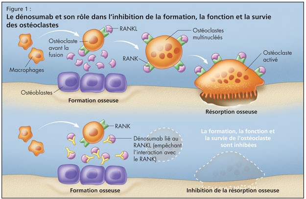
This conundrum was settled when the RANK ligand system was discovered. Receptor activator of nuclear factor-kB ligand (RANKL), a protein that binds to receptor activator of nuclear factor-kB (RANK), is the primary mediator of osteoclast differentiation, activation, and survival (Figure 1). RANK ligand is the primary mediator of bone resorption. Osteoprotegerin (OPG) provides an alternative binding site for RANKL and acts as a decoy receptor by blocking RANK ligand binding to its cellular receptor RANK. Ligand binding activates cellular signalling. Ligand that is bound to a decoy receptor cannot activate cellular signalling.1-6


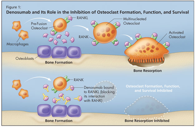
Osteoprotegerin is the natural key endogenous regulator of the RANKL–RANK pathway. When RANK ligand is bound and neutralized by OPG, osteoclasts cannot form,2,5 function,2 or survive.3 Osteoprotegerin acts as a decoy receptor by binding with RANKL, thereby inhibiting osteoclastogenesis and the survival of pre-existing osteoclasts.1,4,7,8 By binding to and neutralizing the effects of RANKL, it thereby inhibits bone resorption.1,4,7,8 More RANKL equates with more bone loss.

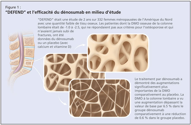
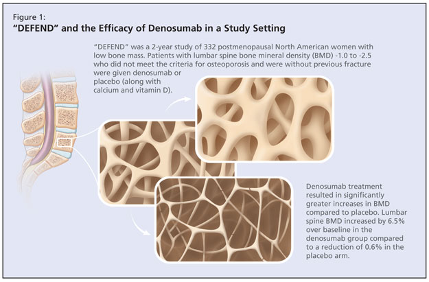
Denosumab is an osteoporosis treatment designed to target RANKL. It is a fully human monoclonal antibody (IgG2) that binds to RANKL with high affinity and specificity. It blocks the interaction of RANKL with RANK, mimicking osteoprotegerin.
The clinical use of OPG is impractical, Dr. Josse observed, and researchers saw the need to create other agents to neutralize RANKL, which increases bone resorption and reduces trabecular and cortical bone. It is implicated in osteoporosis and its inhibition increases bone mineral density and maintains bone architecture. Denosumab is now being investigated in treatments other than osteoporosis, including cancer-related bone destruction and bone erosion in the setting of rheumatoid arthritis.

Dr. Josse reiterated that a healthy skeleton depends on a balance between bone resorption and formation; treatments that are able to achieve RANK ligand inhibition and therefore appropriate suppression of bone resorption could offer significant clinical value to patients with bone loss.1,6

References:

  1. Boyle WJ, Simonet WS, Lacey DL. Osteoclast differentiation and activation. Nature 2003;423:337–42.
  2. Fuller K, Wong B, Fox S, et al. TRANCE is necessary and sufficient for osteoblast-mediated activation of bone resorption in osteoclasts. J Exp Med 1998;188:997–1001.
  3. Lacey DL, Tan HL, Lu J, et al. Osteoprotegerin ligand modulates murine osteoclast survival in vitro and in vivo. Am J Pathol 2000;158:435–48.
  4. Lacey DL, Timms E, Tan HL, et al. Osteoprotegerin ligand is a cytokine that regulates osteoclast differentiation and activation. Cell 1998;93:165–76.
  5. Yasuda H, Shima N, Nakagawa N, et al. Osteoclast differentiation factor is a ligand for osteoprotegerin/osteoclastogenesis-inhibitory factor and is identical to TRANCE/RANKL. Proc Natl Acad Sci USA 1998;95:3597–602.
  6. Hofbauer LC, Schoppet M. Clinical implications of the osteoprotegerin/RANKL/ RANK system for bone and vascular diseases. JAMA 2004;292:490–5.
  7. Simonet WS, Lacey DL, Dunstan CR, et al. Osteoprotegerin: a novel secreted protein involved in the regulation of bone density. Cell 1997;89:309–19.
  8. Bekker PJ, Holloway D, Nakanishi A, et al. The effect of a single dose of osteoprotegerin in postmenopausal women. J Bone Miner Res 2001;16:348–60.

Sponsored by an unrestricted educational grant from Amgen Canada Inc.

Osteoporosis: Preventing the Deterioration of Bone

Osteoporosis: Preventing the Deterioration of Bone

Teaser: 


L. Giangregorio, PhD, Lyndhurst Centre, Toronto Rehabilitation Institute, Toronto, ON.
A. Papaioannou, MD, Department of Medicine, McMaster University, Hamilton, ON.
J.D. Adachi, MD, Department of Medicine, McMaster University, Hamilton, ON.

Osteoporosis is characterized by compromised bone strength, predisposing a person to an increased risk of fracture. The wrist, hip, and spine are the most common sites for fractures associated with osteoporosis. The economic and human costs of osteoporosis-related fractures are considerable. Although it is often considered a woman’s disease, osteoporosis is a significant source of morbidity and mortality in men. Available pharmacological treatments for osteoporosis include bisphosphonates, selective estrogen receptor modulators, calcitonin, parathyroid hormone, and hormone replacement therapy. Non-pharmacological interventions, such as nutritional counselling, exercise, and fall prevention, should also be considered in a fracture prevention plan.
Key words: osteoporosis, fragility fracture, bone, skeleton, bone density.

Building Better Bones

Building Better Bones

Teaser: 

The most effective bone-building treatment may turn out to be a modified form of parathyroid hormone. Researchers report that a peptide formed from the first 34 amino acids of parathyroid hormone is effective in preventing fractures in post-menopausal women. Previously, it was known that injections of parathyroid hormone, or its amino terminal fragment (parathyroid hormone 1-34), increased bone formation and bone mass, but their effects on fractures were unknown.

Women were randomly assigned to receive 20 or 40 mg of parathyroid hormone (1-34) or placebo, administered subcutaneously on a daily basis, for a period of 21 months. Vertebral radiographs and serial measurements of bone mass revealed that the risk of new vertebral fractures and non-vertebral fractures was decreased in the treatment groups; that increases were seen in vertebral, femoral and total-body mineral density; and that the drug is well-tolerated.

The study found that the 40-mg dose increased bone mineral density more than did the 20-mg dose but had similar effects on the risk of fracture and was more likely to have associated side effects.

The study was stopped early because another study on the same peptide fragment found that rats developed bone cancer when given life-long doses. However, researchers eventually decided that the results do not suggest a higher cancer risk in humans.

Source

  1. Neer RM, Arnaud CD, Zanchetta JR, Prince R, Gaich GA, Reginster J et al. New England Journal of Medicine. 2001;344:1434-41.

The Economic Impact of Bone and Joint-Related Health Problems

The Economic Impact of Bone and Joint-Related Health Problems

Teaser: 

A new study by the Institute of Health Economics has estimated that the indirect economic impact of bone and joint problems, including arthritis, totaled $17.9 billion dollars in 1997. Apparently, the main reason for this figure is the lost productivity of people who are unable to work and conduct business. The study reveals that osteoarthritis is two and a half times more prevalent in Canada than is heart disease, and over six times more prevalent than cancer. It is estimated that with Canada's aging population, the number of people with bone and joint-related health problems in Canada will increase by 124% over the next 30 years.

Despite this increased demand, a shortage of orthopaedic surgeons is making it difficult for people to get the care they need. On average, patients are forced to wait more than six months for joint replacement surgery in Canada, and many have to wait longer than a year.

The findings of this study were supported by a parallel phone survey conducted by Decima Research, which found that 42% of Canadians have been affected by bone and joint problems--either personally or through the severe physical pain of a family member or friend.

The Canadian Orthopaedic Association and The Arthritis Society have now developed a comprehensive plan to address the issues that are critical to orthopaedic care in Canada. Entitled Canada in Motion: Mobilizing Access to Orthopaedic Care the plan calls for Canada's federal and provincial ministers of health to work with the medical community to develop a national orthopaedic care strategy.

Please see next month's issue of Geriatrics & Aging for articles on the various options available for the treatment of arthritis in the elderly.

Statin Power for Lowering Lipids and Building Better Bones

Statin Power for Lowering Lipids and Building Better Bones

Teaser: 

Christine Oyugi, BSc
Assistant Managing Editor,
Geriatrics & Aging

Statins are the most effective agents for lowering plasma levels of low-density lipoprotein cholesterol (LDL-C) that are currently available and are the mainstay therapy for the treatment of hyperlipidemia. The drugs are the most commonly prescribed agents for this condition because of their efficacy in reducing LDL, their safety, and their excellent tolerability. Recently, several studies have found that statins also have anabolic effects on bone and may substantially reduce the risk of fractures.1

Mundy et al. were the first to discover the bone anabolic properties of statins.2 Prompted by the observation that bone morphogenic protein 2 (BMP2) causes osteoblasts to proliferate, mature, and form new bone, the researchers screened a library of 30,000 natural compounds to find potential bone strengthening drugs. The study showed that lovastatin (a fungal metabolite); fluvastatin, simvastatin and mevastatin specifically activated the BMP-2 promoter. The researchers also found that oral administration of statins (simvastatin or lovastatin) to rats increased the volume of trabecular bone and the rate of bone formation even in ovariectomized mice.

How Many Bones Must be Broken?

How Many Bones Must be Broken?

Teaser: 

Lilia Malkin, BSc

A fracture is often the first clinical sign of osteoporosis (OP), the silent disease of skeletal fragility characterized by decreased bone mass and deterioration of bone tissue that results in an increased vulnerability to fractures.

The bone mineral density (BMD) criterion frequently used to define osteoporosis was set in 1994 by the World Health Organization (WHO) as more than 2.5 standard deviations below the "young adult mean." An estimated 1.4 million Canadians suffer from OP. In the population aged 50 and over, approximately one in four women and one in eight men are affected. The incidence of the disease increases with age: 70 percent of women have osteoporosis by the age of 80. Predictably, the fracture risk rises with age, with women at higher risk due to both more extensive bone loss and longer average life span. Osteoporotic fractures make a significant contribution to morbidity and mortality in the geriatric population. For instance, the mortality rates within one year of hip fracture are estimated at between 12 and 37 percent, while the average death rate in octogenarians is 2.6 percent per year.

Unfortunately, OP is often asymptomatic prior to the occurrence of a fragility fracture, a break that occurs in the absence of major trauma to the affected bone. The best predictor of fracture risk is low bone density.

The Aging Skeleton--Just the Bare Bones

The Aging Skeleton--Just the Bare Bones

Teaser: 

Rhonda Witte, BSc

Our body's framework is subjected to continual use throughout our daily rituals. Whether we are walking, lifting, exercising or even rolling over in bed while we sleep, we depend on our skeletal system to function adequately. Amazingly, it handles a lot of use and is incredibly reliable. With age, however, our framework becomes less capable of withstanding the "wear and tear" of every day life. Research provides insight into the mechanisms behind the "normal" aging of the skeletal system. With this knowledge, we are gradually learning ways to counteract the effects of aging bone.