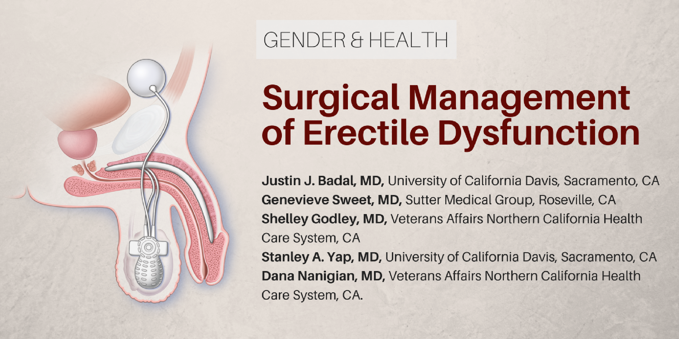
1Department of Urology, University of
California Davis, Sacramento, California.
2Department of Urology, Sutter Medical Group,
Roseville, California.
3Department of Urology, Veterans Affairs
Northern California Health Care System, Sacramento,
California.
4Department of Urology, University of California
Davis, Sacramento, California and Department of Urology,
Veterans Affairs Northern California Health Care System,
Sacramento, California.
5Chief of Urology, Department of Urology,
Veterans Affairs Northern California Health Care System,
Sacramento, California.